
|
Fig. S4
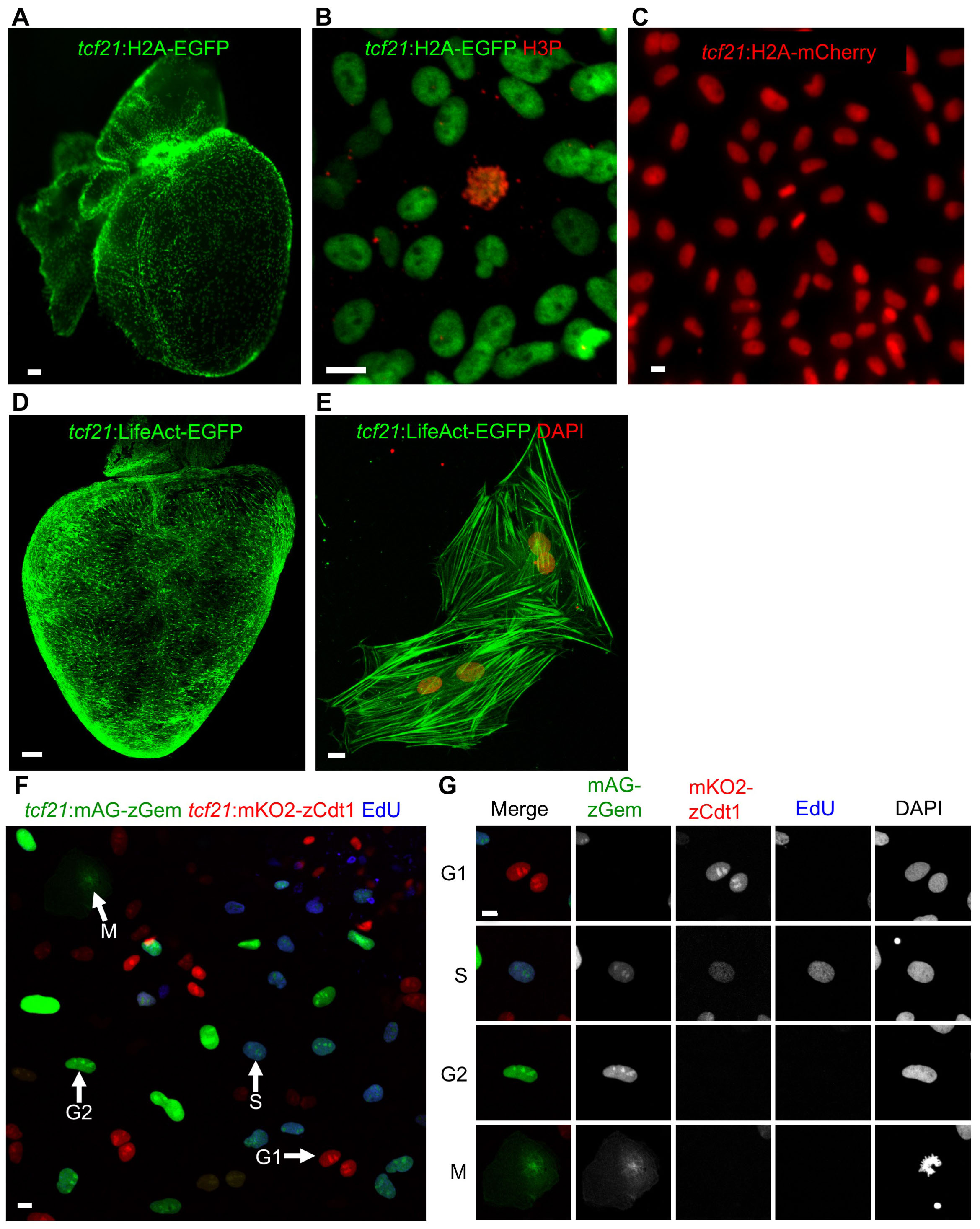
New Reporter Lines for Live Imaging (Related to Figure 3)
(A, B) A tcf21:H2A-EGFP reporter line indicates chromatin in epicardial cells of a wholemounted heart (A) and in epicardial explant culture (B). Anti-phospho-Histone H3 staining (H3P) was performed to label condensed chromatin (red). Scale bars, 100 μm (A) or 10 μm (B).
(C) High-magnification view of tcf21:H2A-mCherry epicardial cells. Scale bar, 10 μm
(D, E) A tcf21:LifeAct-EGFP reporter labels F-actin in whole-mounted (D) or isolated epicardial cells (E). DAPI stains nuclei red in (E). Scale bars, 100 μm (D) or 10 μm (E).
(F, G) Epicardial explant culture from a tcf21:FUCCI heart, stained after a 1 h pulse of EdU to mark S phase. Arrows in (F) denotes different cell cycle phases, also highlighted in (G). Scale bars, 10 μm.
Reprinted from Developmental Cell, 42, Cao, J., Wang, J., Jackman, C.P., Cox, A.H., Trembley, M.A., Balowski, J.J., Cox, B.D., De Simone, A., Dickson, A.L., Di Talia, S., Small, E.M., Kiehart, D.P., Bursac, N., Poss, K.D., Tension Creates an Endoreplication Wavefront that Leads Regeneration of Epicardial Tissue, 600-615.e4, Copyright (2017) with permission from Elsevier. Full text @ Dev. Cell