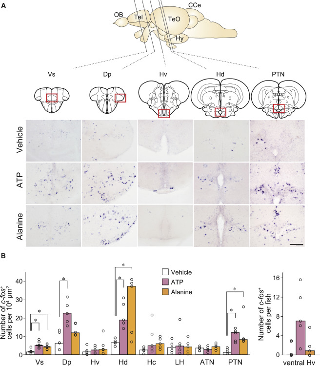
Fig. 3
Higher Brain Centers Activated by ATP and Alanine
(A) In situ hybridization with c-Fos cRNA probe on brain sections of zebrafish exposed to vehicle, ATP, and alanine. Vertical lines in the schematic zebrafish brain indicate the anterior-posterior positions of five coronal sections. Abbreviations for brain regions are as follows: Tel, telencephalon; TeO, optic tectum; Hy, hypothalamus; CCe, cerebellum. Red boxes indicate the locations of magnified views. Scale bar, 100 μm.
(B) Quantification of c-Fos-positive neurons in nine brain regions. Abbreviations for brain regions are as follows: Vs, supracommissural nucleus of ventral telencephalic area; Dp, posterior zone of dorsal telencephalic area; Hv, ventral zone of periventricular hypothalamus; Hd, dorsal zone of periventricular hypothalamus; Hc, central zone of periventricular hypothalamus; LH, lateral hypothalamic nucleus; ATN, anterior tuberal nucleus; PTN, posterior tuberal nucleus. Values represent median (n = 5). Wilcoxon rank sum test with Bonferroni?s correction (Vs, p = 0.024; Dp, p = 0.024; Hd, p = 0.024; PTN, p = 0.024 for vehicle versus ATP; Vs, p = 0.024; Hd, p = 0.048; PTN, p = 0.024 for vehicle versus alanine). ?p < 0.05.

