Fig. S15
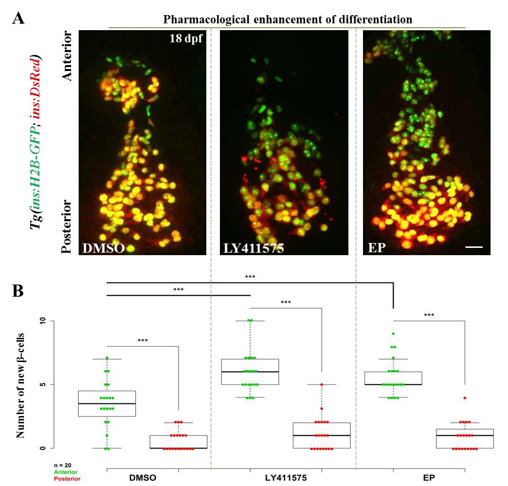
Pharmacological inhibition of Notch and NF-kB signaling increases the number of recently-differentiated cells on the islet?s anterior side. (A) Tg(ins:H2B-GFP);Tg(ins:DsRed) animals were treated with DMSO (control), a ?-secretase inhibitor (LY-411575) or an NF-kB inhibitor (Ethyl Pyruvate (EP)), and the number of recently-differentiated beta-cells was quantified. The recently-differentiated beta-cells are H2B-GFP-positive but DsRed-negative. Maximum intensity projections of 18 dpf primary islets. The anterior side is to the top. (B) Quantification of recently-differentiated beta-cells within the anterior and posterior halves of the primary islet (n = 20 islets) for controls and animals treated with LY-411575 or EP. The plot shows tukey style boxplot overlaid with the data points. In all three cases, the number of recently-differentiated beta-cells was higher within the anterior compared to the posterior half (two-tailed t-test, *** p < 0.001). Treatment with LY-411575 or EP significantly enhanced (two-tailed t-test, *** p < 0.001) the addition of beta-cells within the anterior halves of the islets, as compared to controls, but not within the posterior. Scale bars, 20 ?m.

