Fig. S2
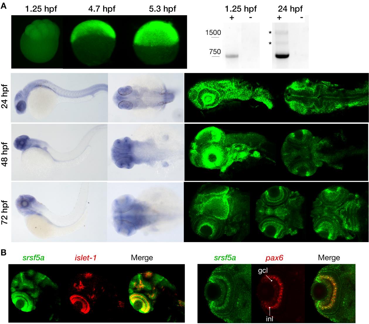
Expression pattern of the srsf5a gene. A, Visualization of maternal and zygotic srsf5a transcripts by whole mount in situ hybridization (ish) (upper left) and RT-PCR analysis (upper right). Maternal expression was detected at 1.25hpf (8 cells). A high expression of srsf5a was observed after zygotic gene activation, as shown by in situ hybridization at 4.7 (30% epiboly) and 5.3hpf (50% epiboly) (lateral views). RT-PCR analysis revealed the presence of two alternative srsf5a transcripts (*) at 24hpf that were absent at 1.25hpf. srsf5a was predominantly expressed in the brain at 24, 48 and 72hpf as determined by visible and fluorescent ish on the bottom right and left, respectively. At 24hpf, we observed an expression in the lens, the optic capsule and in the entire brain, with a more intense signal at the midbrain-hindbrain boundary. At 48hpf, transcripts for srsf5a are detected in the pectoral fins, the otic vesicles, and in the brain with a robust expression in the olfactory bulb, the iris, the optic tectum and in the cerebellum. By 72hpf, the entire brain expresses srsf5a with a more distinct expression in the developing eye, particularly in various layers of the retina, in the optic tectum and in the cerebellum. At this stage, the neuromasts and the pharyngeal region also display srsf5a expression. B, Double fluorescent ish was performed to specify the expression profile in the retina at 72hpf. srsf5a mRNA colocalizes with that of islet-1 and pax6b in the ganglional cell layer (gcl) and in the inner nuclear layer (inl) of the retina.

