Image
Figure Caption
Fig. 3 S1
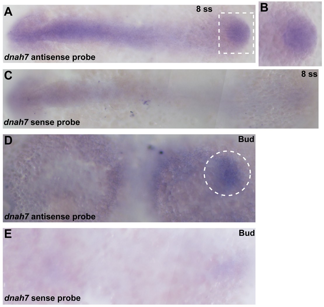
In situ hybridization with dnah7 specific probe in zebrafish embryos.
(A?C) WT 8 ss zebrafish embryo stained with antisense (A?B) and sense (C) dnah7 specific probes. (A) White dotted square delimits the KV, which is detailed in (B). (D?E) WT zebrafish embryos at bud stage, stained with antisense (D) and sense (E) dnah7 specific probes. White dotted line circles the DFCs in (D). In all images Anterior is to left and Posterior is to right.
Acknowledgments
This image is the copyrighted work of the attributed author or publisher, and
ZFIN has permission only to display this image to its users.
Additional permissions should be obtained from the applicable author or publisher of the image.
Full text @ Elife

