
Image
|
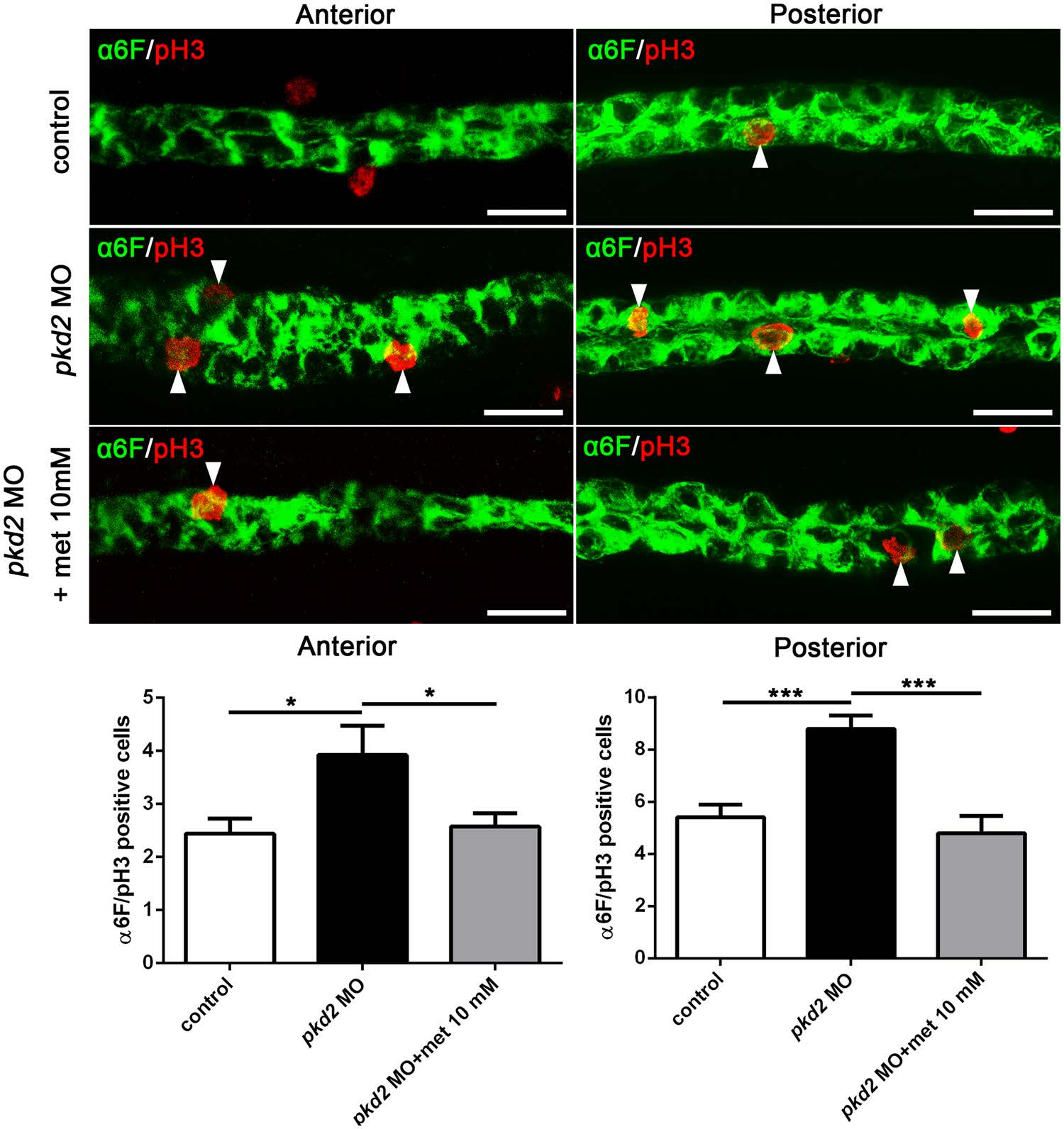
Figure Caption
Fig. 3
Metformin suppresses pronephric epithelial proliferation in pkd2 morphants. (A) Representative confocal immunofluorescence images showing proliferative cells (arrows) in pkd2 morphants with and without metformin (10?mM) treatment. Embryos were stained using anti-PH3 (red) to mark proliferating cells and anti-?6?F (green) to label the pronephric ducts at 48 hpf. (B) Counts of anti-PH3- and anti-?6F-positive cells in the anterior and posterior pronephric ducts (n?=?14?17 per group). *P?<?0.05, ***P?<?0.001. Data represent two independent experiments. Scale bar, 20?�m.
Figure Data
Acknowledgments
This image is the copyrighted work of the attributed author or publisher, and
ZFIN has permission only to display this image to its users.
Additional permissions should be obtained from the applicable author or publisher of the image.
Full text @ Sci. Rep.