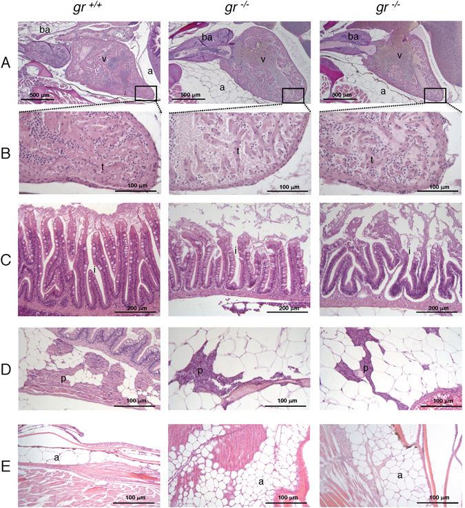
Fig. 2
Histology of 8-month-old gr ?/? and gr +/+ zebrafish. All histological images of 8-month-old gr ?/? and gr +/+ zebrafish were taken from longitudinal sections stained with haematoxylin and eosin (H&E). The left panels represent wild type fish, whereas the middle and right panels show tissues from two mutant fish. (A) heart; (B) particular of the heart at higher magnification showing reduced trabecular network in the mutant samples; (C) intestinal mucosa with sloughing epithelium at the villous tips and reduced height of villi in mutants; (D) visceral view showing reduced extension of pancreas in mutants; (E) consistent increase of subcutaneous adipose tissue in mutants. ba?=?bulbus arteriosus; v?=?ventricle; a?=?adipose tissue; i?=?intestine; p?=?pancreas; t?=?trabeculae.

