Fig. 1
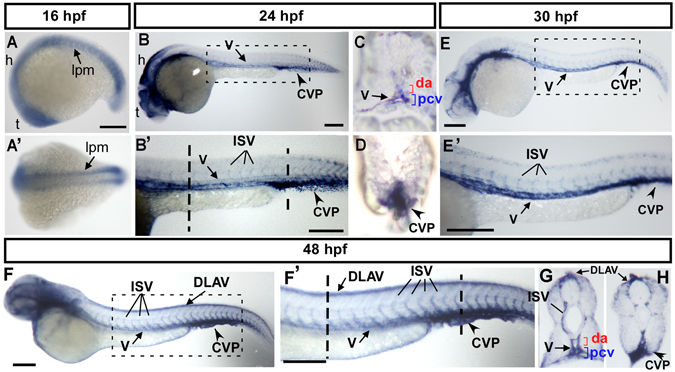
Spatiotemporal expression of cpn1 during zebrafish development. (A) The lateral view shows cpn1 mRNA expression at the 18?S stage in the lateral plate mesoderm (l?pm), hindbrain (h), and telencephalon (t). (A?) The dorsal view of the embryos shows cpn1 expression in the lpm. (B,B?) At 24 hpf, cpn1 was expressed in the vessels (v), intersegmental vessels (ISV) and caudal vein plexus (CVP) of the trunk. B? is an enlargement of B. (C,D) The cross sections of embryos from B? demonstrate that cpn1 is expressed in the dorsal aorta (da), posterior cardinal vein (pcv) and CVP. (E,E?) At 30 hpf, cpn1 was expressed in the vessels (v), ISV, and CVP of the trunk. E? is an enlargement of E. (F,F?,G,H) At 48 hpf, cpn1 was expressed in ISVs, dorsal longitudinal anastomotic vessels (DLAV), vessels (v), da, pcv and CVP, as observed in the lateral view and transverse sections of the embryo trunk and tail regions. F? is an enlargement of F. Scale bars in all figures represent 200?Ám.

