
|
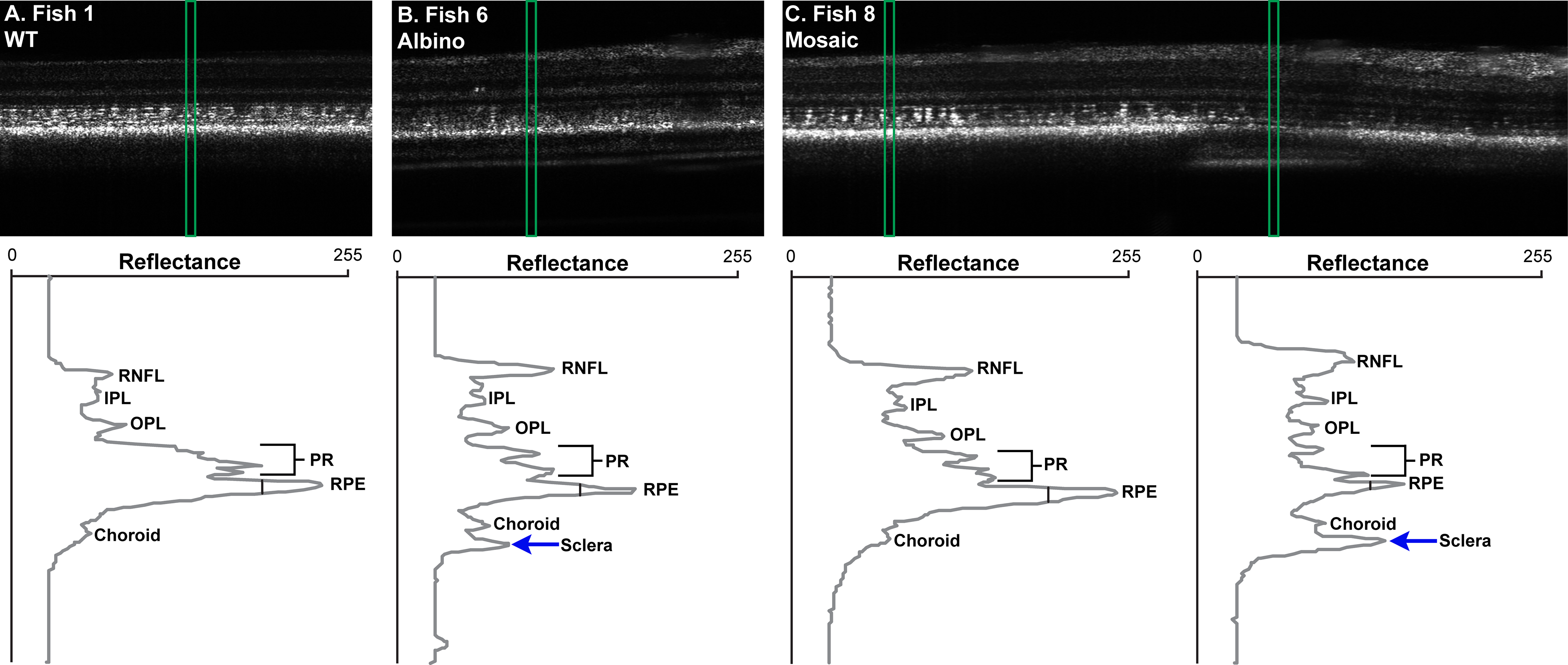
Fig. 4
Differences in LRP peaks with pigmentation in zebrafish. (A) Wild type fish with consistent RPE reflectance and width. (B) Albino fish, whose RPE is distinctly dimmer than the WT. (C) Mosaic fish, where the left LRP was generated in an area with melanin pigment and the right LRP was generated in an area lacking pigment. When melanin in present, the RPE band is wider and has increased reflectance. When melanin is absent, the sclera also is visible posterior to the RPE and choroid. Wild type and albino images are 225 μm wide. Mosaic fish image is 475 μm wide. All images are 479 μm tall. Images are displayed in linear format with intensity values normalized to stretch between 0 and 255 for display only. RNFL, retinal nerve fiber layer; IPL, inner plexiform layer; OPL, outer plexiform layer; PR, photoreceptor layers.