
|
Fig. 3
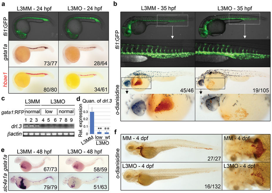
Drl.3 is essential for erythroid development.
(a) Control- (mismatch, L3MM) and drl.3 morpholino- (L3MO) injected embryos at 24?hpf showing live Tg(fli1a:EGFP) marking of the vasculature and gata1a and hbae1 WISH, as indicated. (b) Embryos at 35?hpf showing live Tg(fli1a:EGFP) pattern, with a magnified view of the PBI region, and o-dianisidine stained embryos with an enlargement of the anterior region. (c) RT-PCR analysis of individual Tg(gata1:DsRED) control and L3MO-injected embryos. Drl.3 morphants were sorted for normal or decreased (estimated ? 60% of normal) numbers of circulating erythrocytes at 48?hpf. Full-length gel images for these cropped panels are provided in Supplemental Figure 8. (d) Quantitation of the levels of drl.3 expression normalized to ?-actin from RT-PCR analysis in (c). **P = 0.0019 and **P = 0.0015 (Student's t-test). (e) WISH of gata1a and slc4a1a in 48?hpf control and drl.3 morphants. (f) O-dianisidine stained control and drl.3 morphants at 4?dpf; lateral views (left panels) and ventral views of the anterior region (right panels). (a?b, e?f) The number of the embryos with the representative phenotype per total number of embryos is shown; lateral views with head to the left, dorsal upward.