
Image
|
Figure Caption
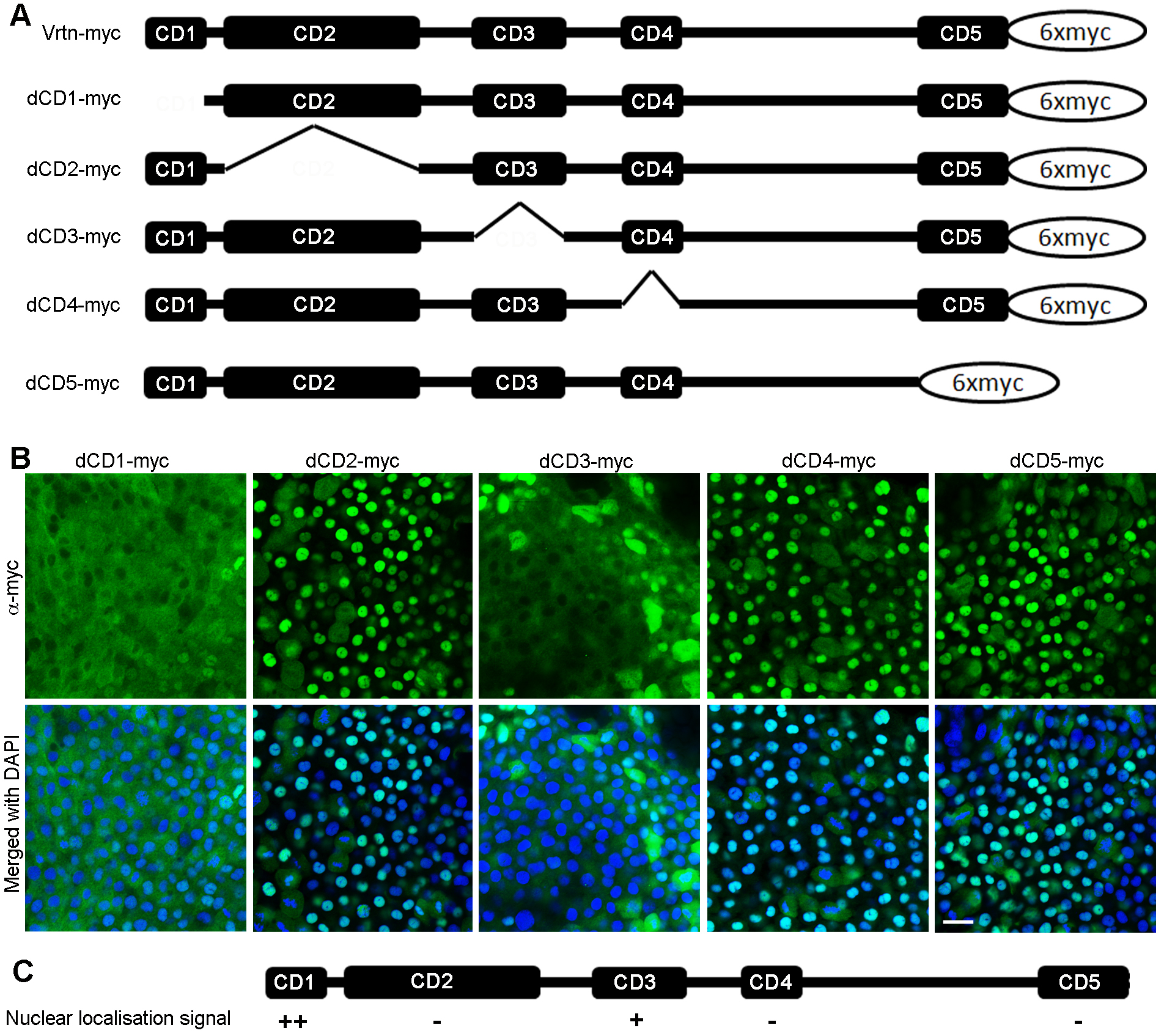
Fig. S15
Subcellular localisation of Vrtn deletion mutants. (A) Diagram showing full-length and truncated forms of Vrtn fused with 6 myc epitopes at the C-terminus. (B) Confocal images showing the distribution of Vrtn deletion mutants in deep cells of embryos at 50% epiboly stage. Note that the nuclear localisation of Vrtn mutants lacking CD1 (dCD1) or CD3 (dCD3) is obviously reduced. (C) Summary of the nuclear localisation activity of different Vrtn deletion mutants. Scale bar: 20 ?m.
Acknowledgments
This image is the copyrighted work of the attributed author or publisher, and
ZFIN has permission only to display this image to its users.
Additional permissions should be obtained from the applicable author or publisher of the image.
Full text @ Development