
|
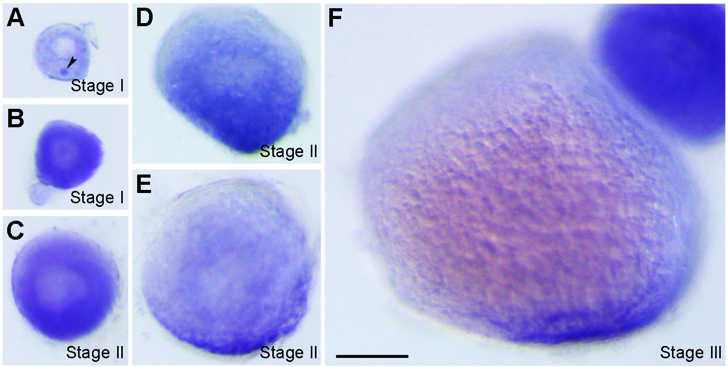
Fig. S7
Localisation of vrtn transcripts during oogenesis. (A) Stage I oocyte (80 ?m in diameter), with vrtn transcripts detected in the ooplasm, and in the Balbiani body (arrowhead). (B) Stage I oocyte (100 ?m in diameter), with strong uniform localisation in the ooplasm. (C) Stage II oocyte (150 ?m in diameter) shows a slight vegetal enrichment. (D) Stage II oocyte (200 ?m in diameter), with evident vegetal localisation. (E) Stage II oocyte (230 ?m in diameter) shows vrtn localisation mainly in the vegetal region. (F) Stage III oocyte with vrtn transcripts restricted exclusively in the vegetal pole cortex. Oocytes were staged according to their diameters: stage I, 70-140 ?m; stage II, 140-340 ?m; stage III, 340-690 ?m. Scale bar: 100 ?m.