Fig. 4 S6
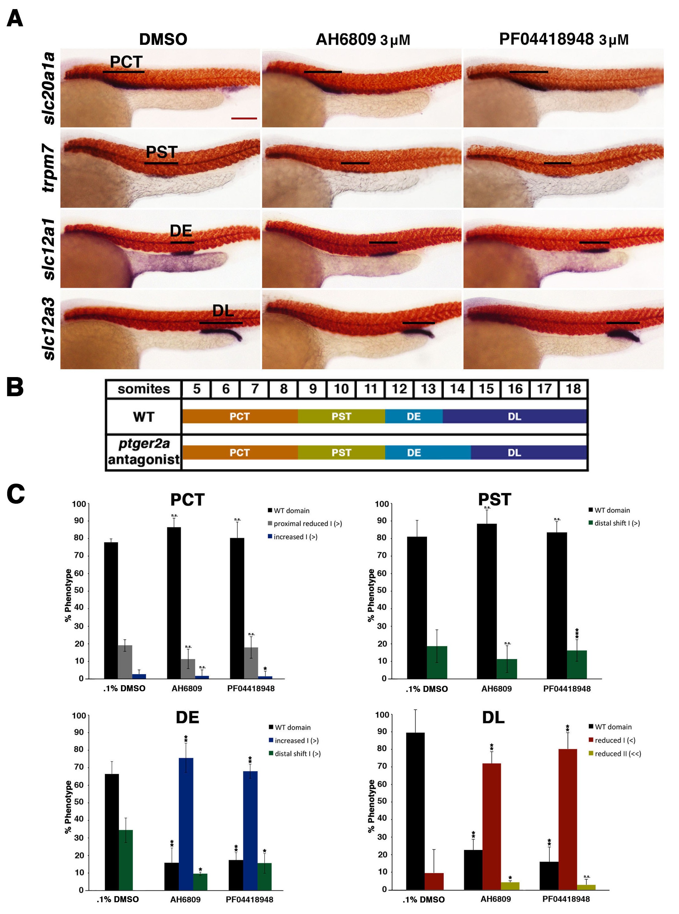
Inhibition of ptger2a using small molecule antagonists promotes distal early fate-choice.
(A) Embryos were exposed to 0.1% DMSO, 3 ?M AH6809, or 3 ?M PF04418948 from 4 hpf to 24 hpf. WISH was used to stain for the PCT (slc20a1a), PST (trpm7), DE (slc12a1), and DL (slc12a3) (purple) and the somites (smyhc1) (red) at the 24 hpf stage. Black bars indicate segment gene expression domain. Red scale bar, 70 Ám. (B) A somite summary schematic describing the changes in patterning caused by ptger2a antagonist treatment. (C) The PCT, PST, DE, and DL segments were quantified per phenotype in triplicate using juxtaposed somites. Greater than (>) and less than (<) were used to categorize embryos, where each (>) or (<) represents the difference of a somite in the pertinent segment area. At least 20 embryos were used for each control and experimental group. Data are represented as ▒ SD significant by t test comparing each drug treatment to the corresponding DMSO control group, *p<0.05, **p<0.005, ***p<0.0005, where n.s. indicates not significant.

