Fig. 1
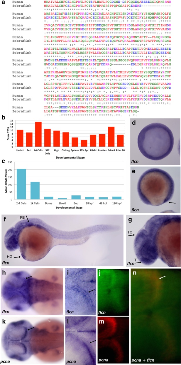
Conservation and expression of zebrafish flcn. a Comparative alignment of human flcn and zebrafish flcn protein sequence using Clustal Omega. (*) indicates positions which have a single, fully conserved residue, (:) indicates conservation between groups of strongly similar properties (.) indicates conservation between groups of weakly similar properties, Red?=?Small (small?+?hydrophobic), Blue?=?Acidic, Magenta?=?Basic ? H, Green?=?Hydroxyl?+?sulfhydryl?+?amine?+?G and Grey?=?Unusual amino/imino acids. b Graph showing CAGE CT values for the zebrafish flcn gene over time, where CT values are the sum of all CAGE detected Transcription Start Site (CTSS) values , representing the number of cage tags (initiation instances) occurring at the same base (normalized tags per million (tps)) in the flcn promoter region. The count value was normalized according to total mapped tags and CTSS instances. c Graph showing the RNAseq RPKM values for zebrafish flcn over time, where the RPKM values represent the mean of the signal coverage value in the promoter region, therefore the sum of the total signal in the whole gene locus region divided by the number of bases with coverage. d Shield stage embryo showing ubiquitous expression. e 4 somite stage embryo showing low level expression over the whole embryo with increased expression seen in the region of the tail bud (arrow). f Prim-10 stage embryo showing low level expression in the whole embryo with more pronounced expression in the forebrain and hindbrain areas, hatching gland (HG, arrow) and the fin bud (FB, arrow). g Head of Prim-10 stage embryo showing expression in the tectum (TC) and telencephalon (T) (arrows). h Long-pec stage embryos showing expression in the brain (arrow) and retina of the embryo. i Long-pec stage embryo showing pronounced expression in the posterior tectum of the brain (arrow). j Head region of a Long-pec stage embryo showing expression of flcn (green) in the posterior tectum. k Long-pec stage embryo showing pcna expression in the retina and brain (arrow). l pcna expression in the posterior tectum of a Long-pec stage embryo (arrow) (m) head region of the same Long-pec stage embryo as panel J showing pcna (red) in the posterior tectum (n) head region of a Long-pec stage embryo showing expression of both pcna (red) and flcn (green) with co-localisation (yellow) in the posterior tectum (n?=?20 embryos for all WMISH)

