
|
Fig. 2
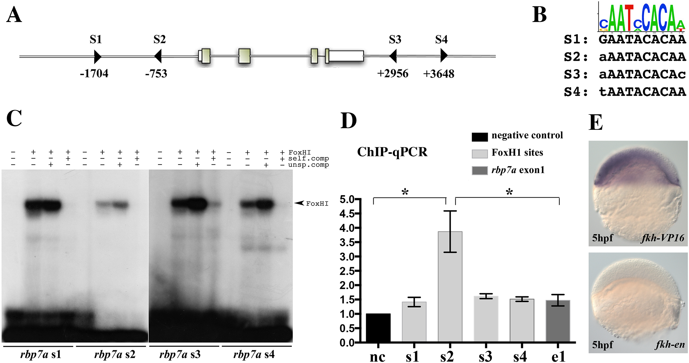
rbp7a is a direct target of FoxH1.
[A] Schematic drawing of the rbp7a gene highlighting 4 potential FoxH1 binding sites (triangles: S1, S2, S3, and S4). Exons are indicated by boxes, the numbers correspond to the distance form the transcriptional start site. [B] Sequence comparison of the mouse FoxH1 consensus log [37] with the four potential sites in rbp7a. [C] In vitro EMSA studies with translated FoxH1 protein with oligonucleotides containing the four potential FoxH1 binding sites (sequences and positions were shown in [A, B]). Competition experiments with unspecific (unsp.comp) and specific (self.comp) unlabeled oligonucleotides were added to verify the binding specificity. [D] In vivo chromatin immunoprecipitation (ChIP-qPCR) experiments performed with 6hpf eGFP-foxH1 mRNA injected MZsur embryos. Bars show the enrichments of DNA fragments in the regions of FoxH1 binding sites in relation to a negative control region (rhodopsin promoter region) that was lacking FoxH1 binding sites (*P<0.05; error bars indicated the SEM). [E] Induced and depleted rbp7a expression in 5hpf wild type embryos after injection of fkh-vp16 and fkh-en mRNA, respectively.