
|
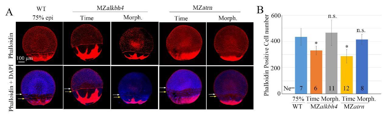
Fig. S1
EVL and deep cell movements are defective in MZalkbh4 and MZatrn embryos, while the cell number per embryo is not obviously affected. (A) Whole-mount phalloidin and DAPI staining were used to observe the marginal positions of EVL and deep cells. White arrows indicate the marginal positions of deep cells; yellow arrows indicate the marginal positions of EVL cells. The mutant embryos were collected at both the same time point and comparable morphological stages compared with wild-type embryos at 75% epiboly stage. (B) The quantitative data of phalloidin-positive EVL cell numbers were derived from 3D images of the embryos in (A). Ne, the number of observed embryos. *, p<0.01; n.s. indicates no significant difference. Scale bars 100 µm in (A).