
|
Fig. S6
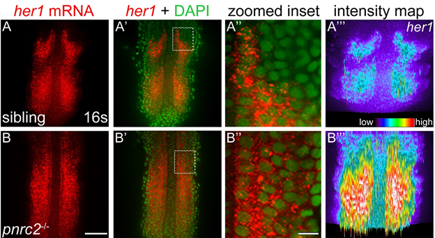
her1 mRNA accumulates dramatically in pnrc2oz22 mutants. Detection of her1 mRNA by in situ hybridization chain reaction (HCR-ISH) reveals her1 misexpression throughout the presomitic mesoderm (PSM) of pnrc2 mutants (A, B). Representative embryos are shown (n=5 in A, n=5 in B). Substantial cytoplasmic localization revealed with DAPI counter staining is apparent in both unaffected siblings and pnrc2 mutants (A', B', 470X magnified in A”, B”). Because detection of her1 mRNA in pnrc2oz22 mutants is very high relative to wild type, levels have been reduced in pnrc2 mutant panels (B-B”). When wild-type and pnrc2 mutants are imaged at the same laser level, raw intensity maps of her1 HCR-ISH signal reveal the extent of her1 mRNA accumulation in pnrc2oz22 mutants (A''', B'''). Scale bars = 50 um (B), 50 nm (B'').
Reprinted from Developmental Biology, 429(1), Gallagher, T.L., Tietz, K.T., Morrow, Z.T., McCammon, J.M., Goldrich, M.L., Derr, N.L., Amacher, S.L., Pnrc2 regulates 3'UTR-mediated decay of segmentation clock-associated transcripts during zebrafish segmentation, 225-239, Copyright (2017) with permission from Elsevier. Full text @ Dev. Biol.