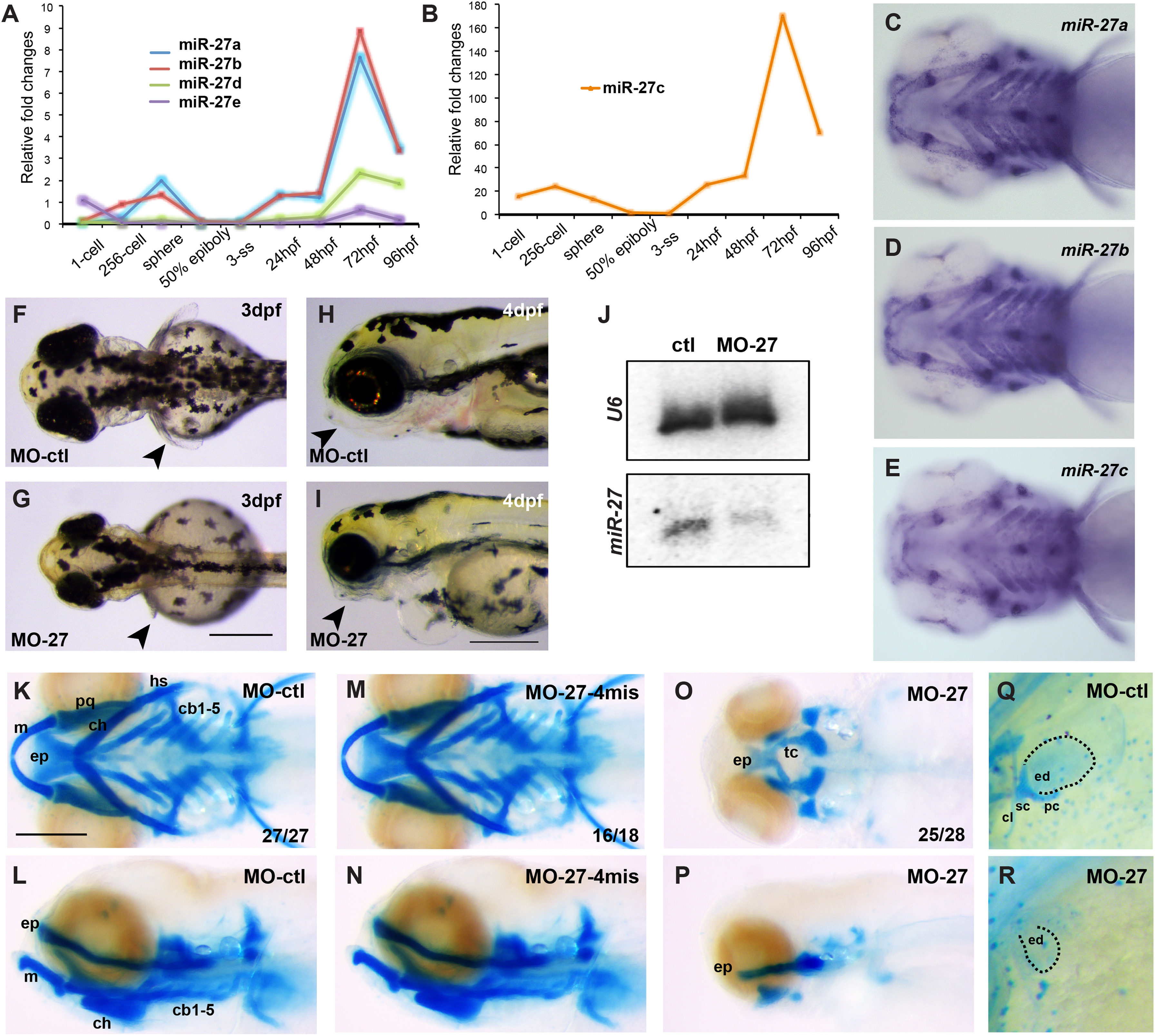
Fig. 1
Knock down of miR-27 leads to craniofacial and pectoral fin defects. (A,B) qRT-PCR for miR-27a-e at the indicated developmental stages normalized to U6 snRNA. Fold changes were calculated using ??C(t) method comparing all miR-27 levels to miR-27c levels at the 3 somite stage (ss). Due to comparably higher levels, miR-27cexpression profile is shown separately. (C-E) Expression of miR-27a, miR-27b and miR-27c in 4dpf embryos detected by whole-mount in situ hybridization by locked nucleic acid (LNA) probes. All are ventral views of the head. (F,G) Dorsal view of 3 dpf live embryos injected with either 5 ng standard control morpholino (MO-ctl) or MO-27 at the single-cell stage. Pectoral fins are indicated with arrowheads. (H,I) Morphology of the head in 4 dpf embryos injected with either MO-ctl or MO-27. Lateral views, jaws are indicated with arrowheads. Scale bar, 300 Ám. (J) miR-27 and U6 levels in uninjected control and miR-27 morpholino (MO-27) injected embryos at 48hpf detected by Northern blot. (K-P) Head cartilages stained with Alcian blue in 4dpf embryos injected with (K,L) standard control morpholino (MO-ctl), (M,N) 4-mismatch miR-27 morpholino (MO27-4mis) and (O,P) MO-27. Top panels, ventral views; bottom panels, lateral views. The indicated ratio represents the number of embryos with the represented phenotype/total number of observed embryos. Cartilage labels: ep, ethmoid plate; tc, trabeculae cranii; m, Meckel's cartilage; pq, palatoquadrate; ch, ceratohyal; hs, hyosymplectic; cb, ceratobranchial. Anterior side of the embryos is to the left. (Q,R) Staining of pectoral fin skeleton in 4dpf embryos by Alcian blue. The right side pectoral fin is shown with anterior to the top. The cleithrum (cl) and scapulocoracoid (sc) cartilages and postcoracoid process (pc) of pectoral fins are missing and the endoskeletal disc cartilage (ed) is smaller in miR-27morphants compared to the controls. Scale bar, 200 Ám.
Reprinted from Developmental Biology, 429(1), Kara, N., Wei, C., Commanday, A.C., Patton, J.G., miR-27 regulates chondrogenesis by suppressing Focal Adhesion Kinase during pharyngeal arch development, 321-334, Copyright (2017) with permission from Elsevier. Full text @ Dev. Biol.

