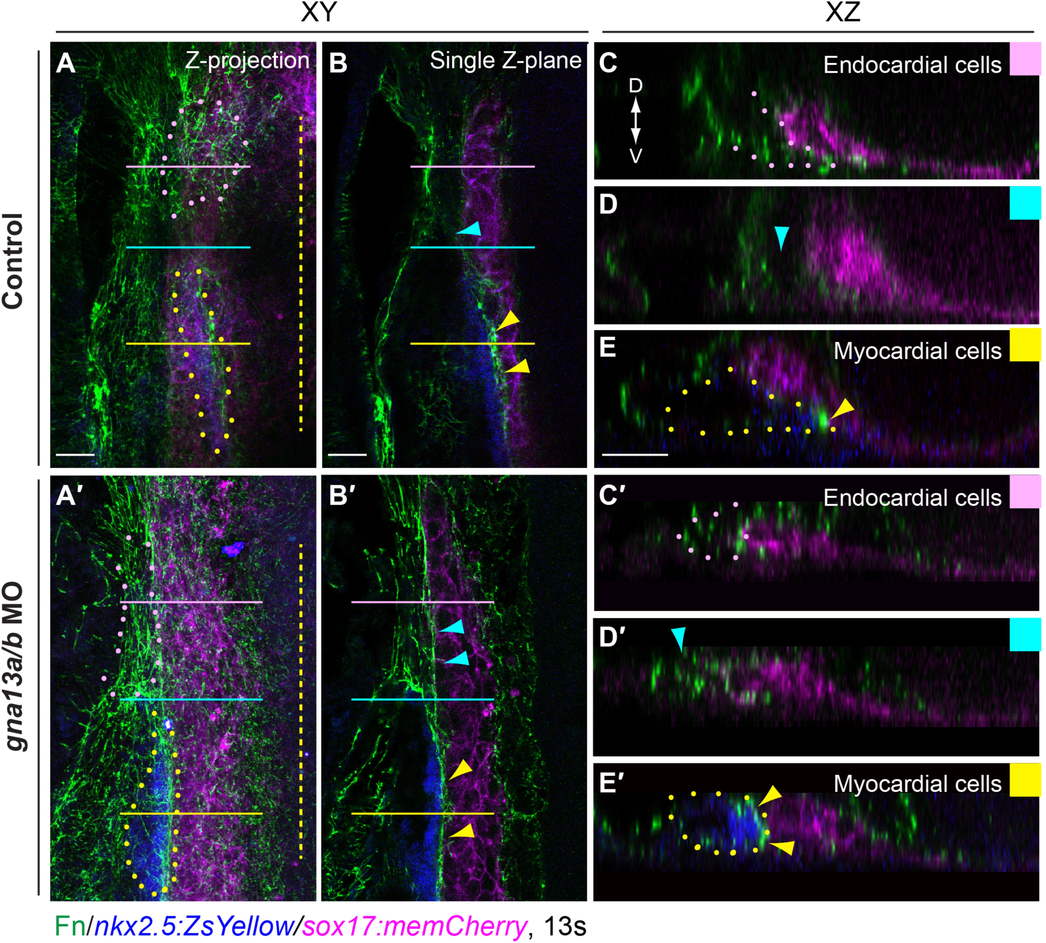
Fig. 7
In G?13 morphants, Fn fibrils in the regions in which endocardial and myocardial precursors reside are disrupted. Whole-mount Fn immunostaining was performed in control (A-E, n=6) and gna13a/b MO-injected (A?-E?, n=7) Tg(nkx2.5: ZsYellow/sox17: memCherry) embryos at 13 s (A, A?) Projections of XY views of confocal Z-stacks spanning the left-hand side of the embryo, with labeled myocardial precursors (blue), endoderm (magenta), and Fn fibrils (green). (B, B?) A single Z-plane from Z-stacks in A, A?. (C-E, C?-E?) Images of XZ transverse sections (corresponding colored boxes on the far right corners) of the regions indicated by horizontal pink, yellow and cyan lines in A, A?. Pink dots: presumptive endocardial cell populations; yellow dots: myocardial cell populations; pink arrowheads: leading regions of myocardial populations; yellow arrowheads: regions between endocardial and myocardial precursors; yellow dashed line: midline; D: dorsal; V: ventral. Scale bars: 20 Ám.
Reprinted from Developmental Biology, 414, Xie, H., Ye, D., Sepich, D., Lin, F., S1pr2/G?13 signaling regulates the migration of endocardial precursors by controlling endoderm convergence, 228-43, Copyright (2016) with permission from Elsevier. Full text @ Dev. Biol.

