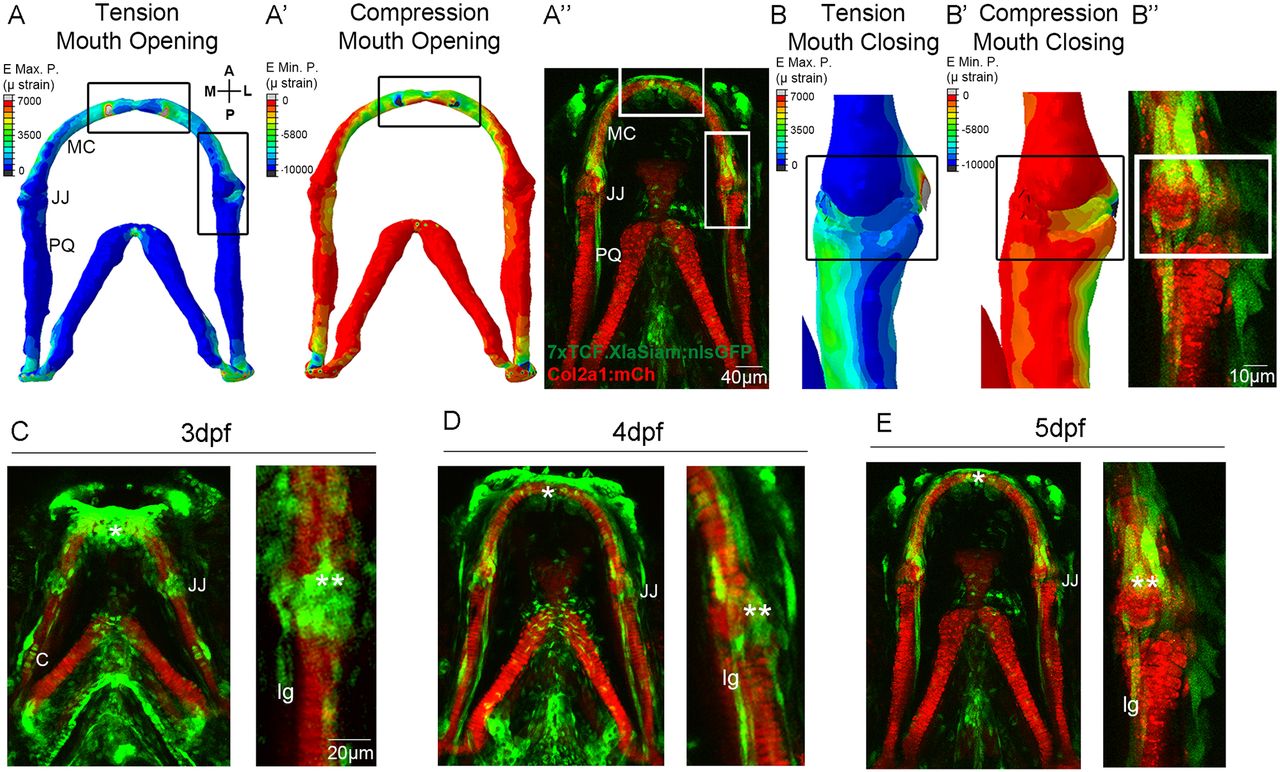
Fig. 1
Patterns of biomechanical strain and the location of Wnt-responsive cells at the zebrafish lower jaw between 3 and 5 dpf. (A,A?) Finite element (FE) model of maximum (E max. P., tension, A) and minimum (E min. P., compression, A?) principal strain on the zebrafish lower jaw during mouth opening at 5?dpf. (B,B?) FE model of maximum (E max. P., tension, B) and minimum (E min. P., compression, B?) principal strain on the jaw joint during mouth closure at 5?dpf. Colour key represents strain in microstrain (Ástrain units). (A?,B?,C-E) Tg(7xTCF.XlaSiam:nlsGFP) and Tg(Col2a1aBAC:mcherry) transgenic zebrafish lines with, respectively, the Wnt-responsive cells (green) and cartilage (red) of the lower jaw labelled at 3 (C), 4 (D) and 5?dpf (A?,B?,E). (C-E) Left: lower jaw. Right and B?: jaw joint. (A,A?,B,B?) Reproduced fromBrunt et al. (2015), where it was published under a CC-BY license (https://creativecommons.org/licenses/by/4.0/). A, anterior; P, posterior; M, medial; L, lateral; MC, Meckel's cartilage; JJ, jaw joint; PQ, palatoquadrate; C, cartilage; lg, ligament; *anterior MC; **jaw joint. Scale bars: 40 ?m in A?; 10 ?m in B?; 20 ?m in C-E.

