
|
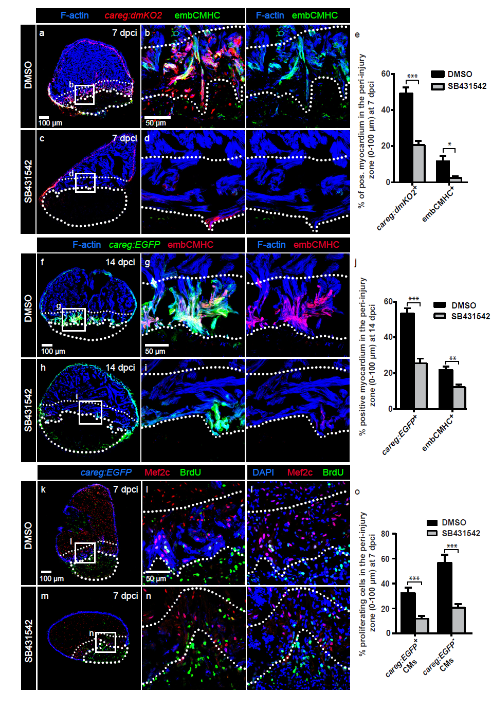
Fig. S7
The inhibition of TGF?/Activin-? signaling suppresses CM dedifferentiation and proliferation at the injury-abutting zone during heart regeneration.
(a-d, f-i) Immunofluorescence staining of ventricles at 7 dpci (a-d) or 14 dpci (f-i) treated with DMSO or 20 ?M SB431542 labelled with antibodies against fluorescent proteins and embCMHC. The intact myocardium is detected by F-actin staining (blue). The upper dotted lines indicate the 100 ?m-thick margin of the myocardium along the injury border. (e, j) Percentage of positive myocardium for the careg reporter or embCMHC in a 100 ?m-wide margin of the regenerating myocardium in control or SB431542-treated hearts. N ? 5. ***p < 0.0001, **p < 0.01, *p < 0.01; unpaired t-test. Error bars correspond to standard error of the mean (SEM). (k-n) Immunofluorescence staining of careg:EGFP ventricle at 7 dpci treated with DMSO or 20 ?M SB431542 labelled with antibodies against GFP (blue), Mef2c (cardiac nuclei, red) and BrdU (green). (o) Percentage of BrdU+ CMs in a 100 ?m-wide margin of the regenerating myocardium in control or SB431542-treated hearts. N ? 5. ***p < 0.0001; unpaired t-test. Error bars correspond to standard error of the mean (SEM).