
|
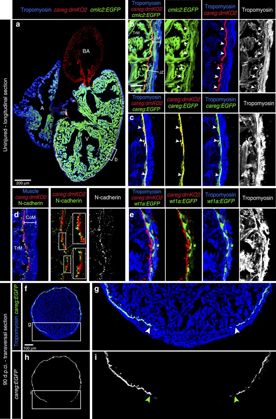
Fig. 8
Monitoring the primordial CM layer between the compact and trabecular myocardium.
(a) Longitudinal sections of careg:dmKO2;cmlc2:EGFP transgenic adult hearts labelled with antibodies against KO2 (red) and Tropomyosin (blue, white). V, ventricle; A, atrium; BA, bulbus arteriosus. (b) careg:dmKO2 expression is maintained during adulthood in a single layer of thin subcortical CMs (arrowheads) in the junctional zone (JZ) between the compact/cortical (CoM) and the trabecular myocardium (TrM). N=4. (c) A higher magnification of the adult ventricle wall of double transgenic fish careg:dmKO2; careg:EGFP displays an overlapping expression of both markers in subcortical myocytes (arrowheads). N=4. (d) A magnified adult ventricle wall of careg:dmKO2 transgenic fish displays an abundant expression of N-cadherin (green, white) on the surface of the junctional CMs on the side facing the trabecular myocardium. N=4. (e) A fragment of the adult ventricle wall of careg:dmKO2;wt1a-6.8kb:GFP transgenic fish displays the separation of compact myocardium and subcortical careg:dmKO2+ layer by wt1a-6.8kb:GFP-labelled fibroblasts of the junctional zone. N=4. (f?i) At 90 d.p.ci., transversal heart sections of careg:EGFP hearts display a gap in the primordial layer within the regenerated myocardium. The extent of the gap is indicated with arrowheads. N=4.