
|
Fig. 6
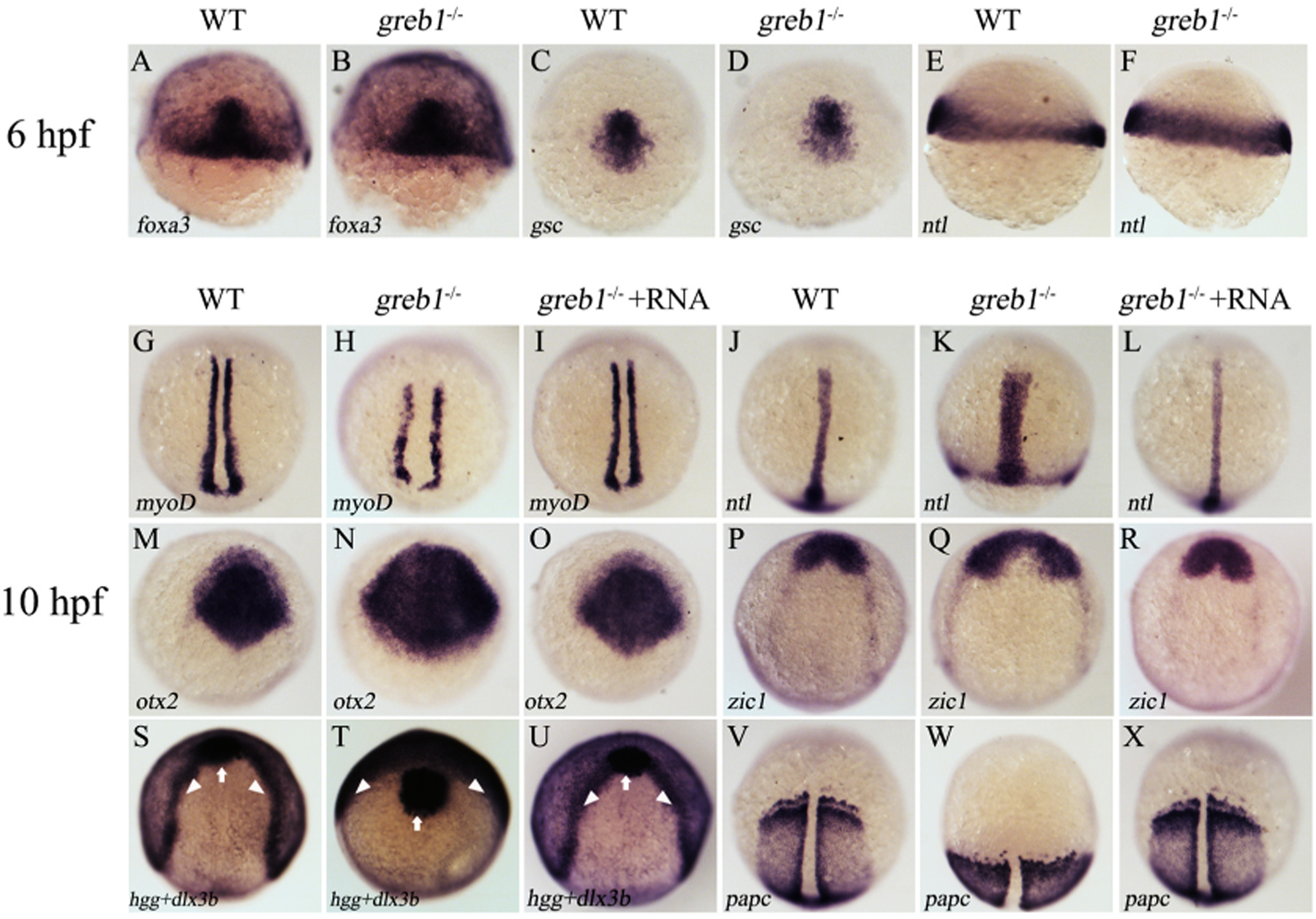
Whole mount in situ hybridization detection of different marker genes in WT, greb1?/? and rescued (greb1?/? + RNA) embryos at 6 hpf and 10 hpf. A and B, foxa3; C and D, gsc; E and F, ntl; G?I, myoD (staining the adaxial cell); J?L, ntl (staining the forerunner cell group, axial chorda mesoderm); M?O, otx2 (staining the anterior axial hypoblast and neural plate); P?R, zic1 (staining the neural plate); S?U, dlx3b (white arrowheads) and hgg (staining the prechordal plate, indicated by white arrows); V?X, papc (staining the paraxial mesoderm). Embryos in panels A?F were shown in lateral view with dorsal side on the right, and embryos in panels G?X were shown in dorsal views with the anterior to the top.
Reprinted from Gene, 627, Li, S.Z., Liu, W., Li, Z., Li, W.H., Wang, Y., Zhou, L., Gui, J.F., greb1 regulates convergent extension movement and pituitary development in zebrafish, 176-187, Copyright (2017) with permission from Elsevier. Full text @ Gene