
Image
|
Figure Caption
Fig. 3
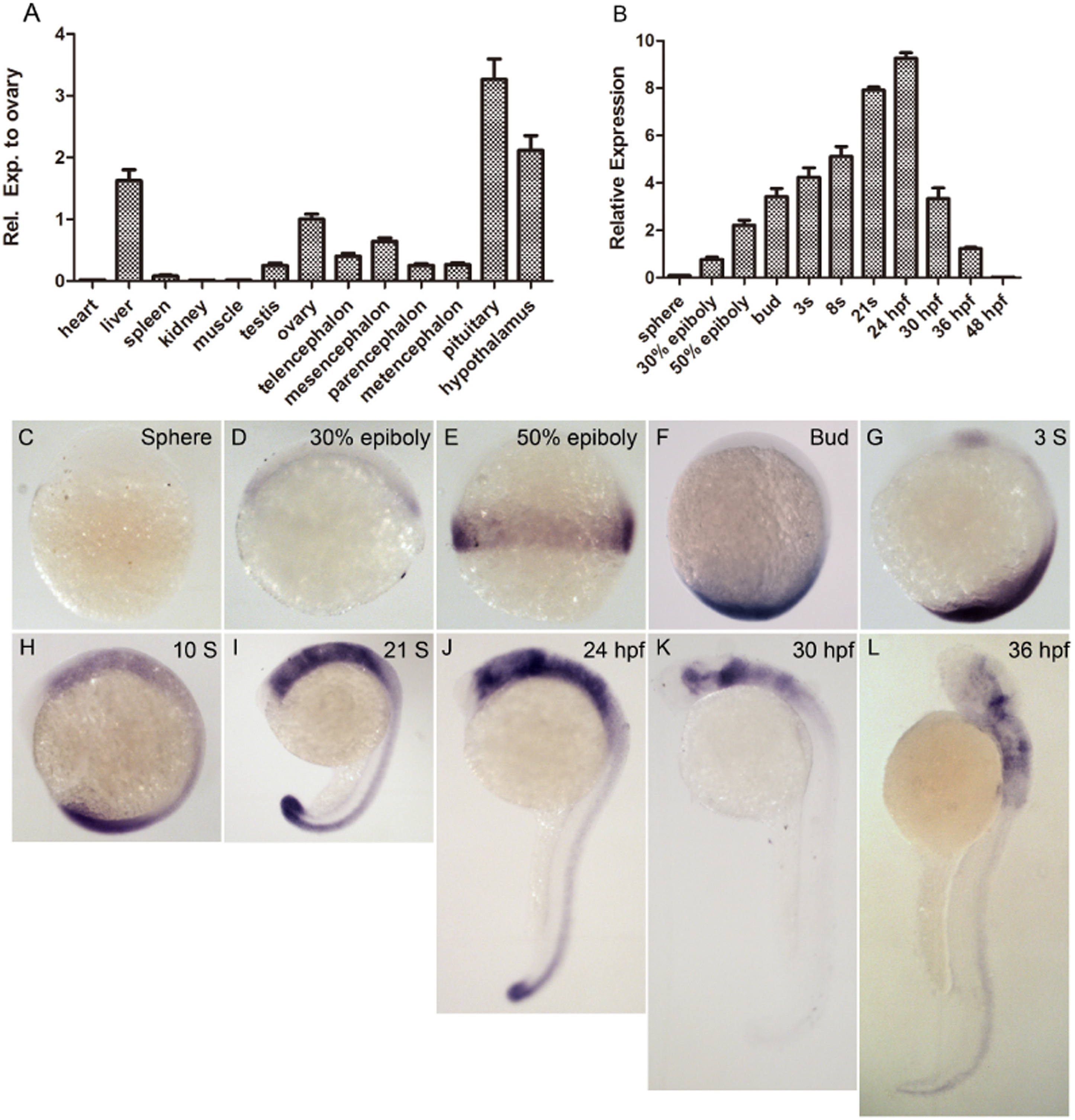
Expression patterns of greb1 in tissues and embryos. (A, B) Real time PCR detection of greb1 transcripts in adult tissues (A) and embryos of different developmental stages (B). (C?L) Whole mount in situ hybridization of greb1 transcript in embryos at different development stages including (C) sphere, (D) 30% epiboly, (E) shield, (F) bud, (G) 3S (3-somite), (H) 10S (10-somite), (I) 21S (21-somite), (J) 24 hpf (prim-5), (K) 30 hpf (prim-15) and (L) 36 hpf (prim-23). The embryos are lateral views with animal pole toward the top and dorsal to the right.
Figure Data
Acknowledgments
This image is the copyrighted work of the attributed author or publisher, and
ZFIN has permission only to display this image to its users.
Additional permissions should be obtained from the applicable author or publisher of the image.
Reprinted from Gene, 627, Li, S.Z., Liu, W., Li, Z., Li, W.H., Wang, Y., Zhou, L., Gui, J.F., greb1 regulates convergent extension movement and pituitary development in zebrafish, 176-187, Copyright (2017) with permission from Elsevier. Full text @ Gene