
|
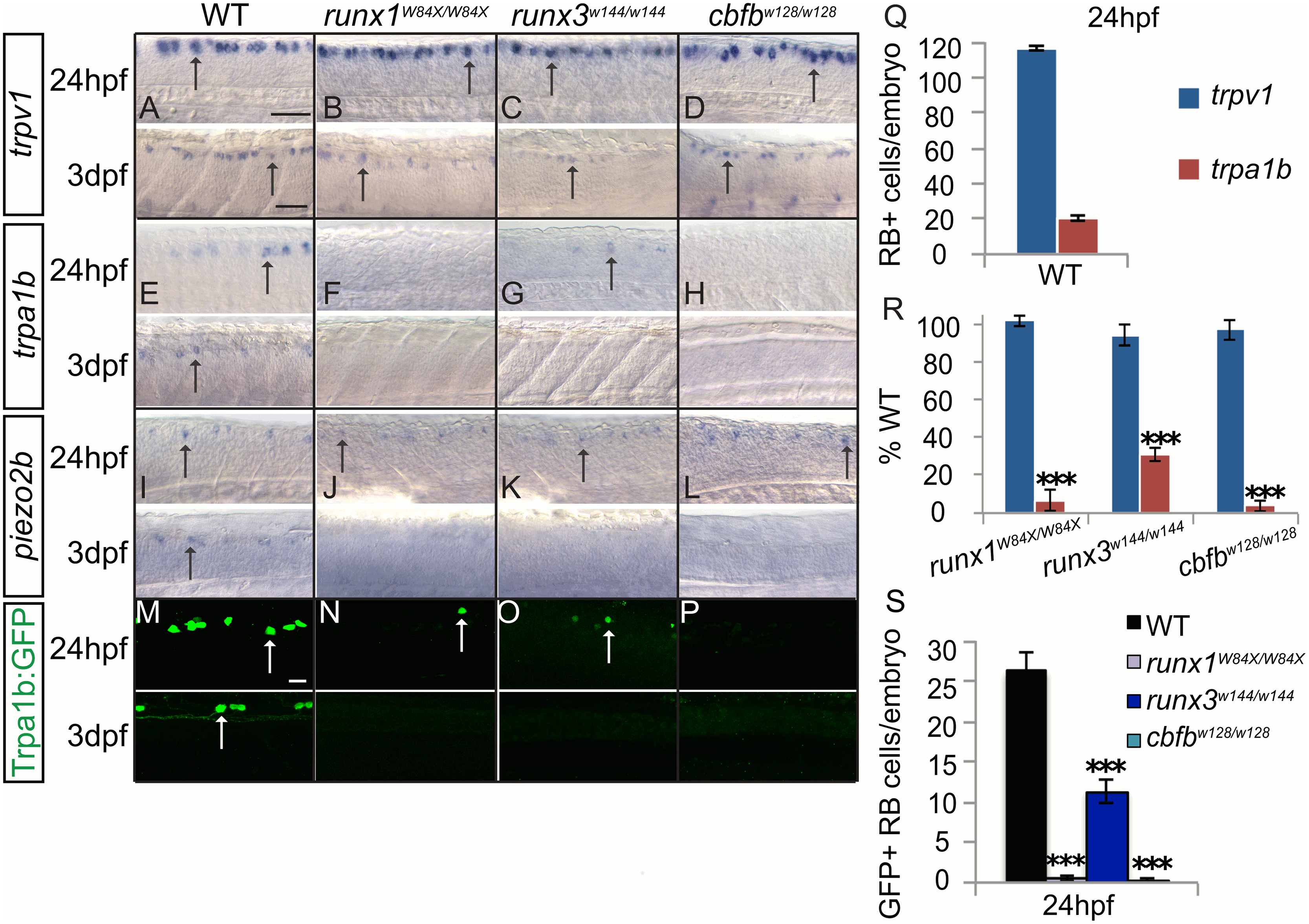
Fig. 8
Loss of runx or cbfb expression affects ion channel expression in the RBs.
A-L, Colormetric in situ hybridization for trpv1 (A-D), trpa1b (E-H), and piezo2b (I-L) in the runx1W84X/W84X, runx3w144/w144 and cbfbw128/w128 mutants focusing on the RBs at 24hpf and 3dpf. M-P, Antibody staining of GFP (green) in runx3w144/w144; trpa1b:GFP and cbfbw128/w128; trpa1b:GFP mutants. Q-R, Quantification of marker gene expression as total number of RB neurons/embryo and as % WT at 24hpf. S, Quantification of GFP+ cells in the RBs at 24hpf. Dashed line outlines the eye; Arrow, RBs; Arrowhead, TG; X, vagal ganglion. Scale bar: A-L 100 ?m, J-L, M-N 20?m. Embryos per condition (n = 3?12). ***p<0.001. All error bars represent S.E.M.