
Image
|
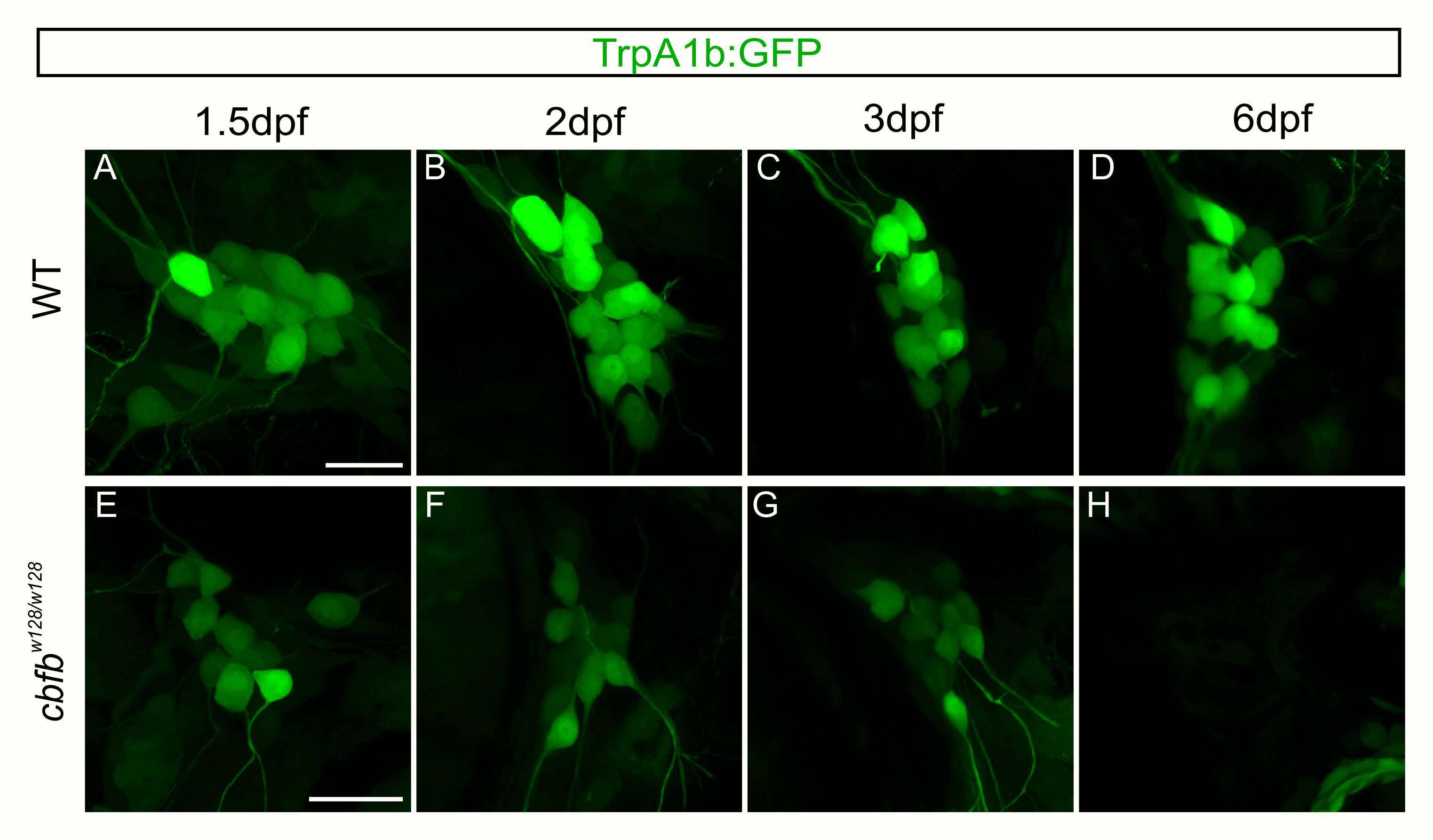
Figure Caption
Fig. S8
Residual GFP allows for visualization of TrpA1b fated neurons in the in the cbfbw128/w128 mutant.
A-H. Maximum intensity projections of TrpA1b:GFP expression from the same TG in a WT (A- D) or cbfbw128/w128 (E-H) embryo over five days. Scale bar: 20μm.
Acknowledgments
This image is the copyrighted work of the attributed author or publisher, and
ZFIN has permission only to display this image to its users.
Additional permissions should be obtained from the applicable author or publisher of the image.
Full text @ PLoS Genet.