Fig. 6
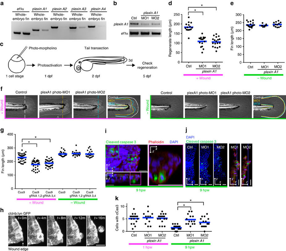
Plexin A1 regulates wound repair in zebrafish fins.
(a) RT?PCR of plexins. plexin A1 and A3 are relatively enriched in tail fins at 2 d.p.f. (b) RT?PCR of plexin A1 following knockdown. Two independent splice morpholinos were used. (c) Diagram of photo-morpholino injection, photoactivation and tail transection. (d) Regenerated fin length at 3 days post wounding (d.p.w.), i.e., 5 d.p.f. *P<0.05; one-way ANOVA with Dunnett?s post-test. (e) Fin length at 5 d.p.f. in the absence of wounding. (f) Images of 5 d.p.f.-larval tail fins with/without wounding. Note that wounding affects both the length and width of the wounded tail fins in plexin A1 morphants. (g) CRISPR-mediated plexin A1 inhibition impairs fin regeneration at 3 d.p.w. (5 d.p.f.) but not the fin length without wounding. *P<0.05; one-way ANOVA with Dunnett?s post-test. (h) Epithelial cell extrusion around a wound of Tg(cldnb:lynGFP). On average, 2.75 (n=4, s.e.m.=0.48) cells were observed to be extruded for 5?h imaging, but note that all the extrusion events were not captured because imaging was done only in a part of the tail fin. (i) An extruded cell is undergoing apoptosis. (j) Representative images of transected tail fins at 9?h.p.w. (k) Knockdown of Plexin A1 increases apoptotic cells at wounds at 9?h.p.w. but not 1?h.p.w. *P<0.05; one-way ANOVA with Dunnett?s post-test.

