Fig. S2
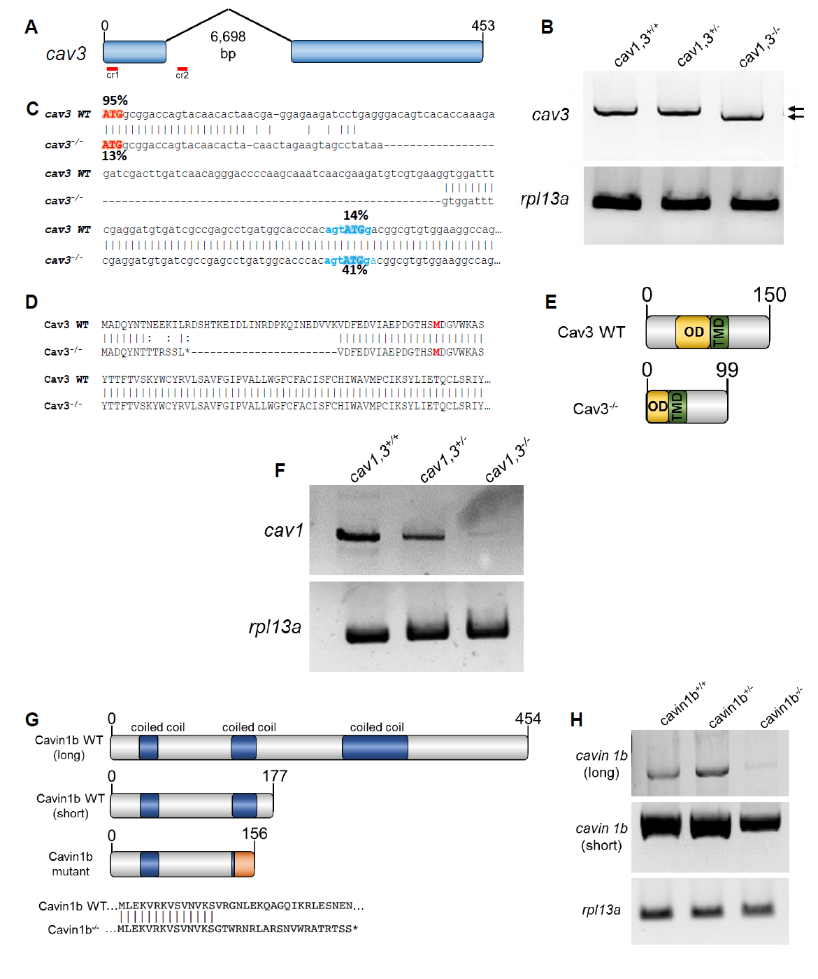
Generation and molecular characterization of cav3 and cavin1b mutants. Related to Figure 1 and 2.
A: Generation of cav3pd1149 allele. Two guide RNAs were injected simultaneously to delete a genomic region spanning most of exon one and part of the first intron.
B: RT-PCR using primers pairing in the 5? and 3? UTR of cav3 revealed that the cav3pd1149 is spliced to produce a slightly shorter transcript. WT and mutant transcripts are marked by arrows rpl13a was used as standard.
C: cDNA sequence of WT and cav3pd1149 mutant transcripts. The % next to each ATG codon indicates the probability of functioning as a start site. The Kozac sequence of the alternative start site is indicated in blue.
D: Predicted protein sequence produced by the WT and cav3pd1149 mutant transcripts. The genomic deletion creates an early stop codon and a deletion of part of the N-terminus in the mutant. The alternative first aa is indicated in bold.
E: Domain structure of WT and mutant Cav3. The N-terminus and part of the oligomerization domain (OD) are eliminated, leaving the trans-membrane domain (TMD) and the C-terminus intact.
F: RT-PCR of ?/? cav1. Both cav1 transcripts largely decay in cav1, 3 mutants. rpl13a was used as standard.
G: Domain structure of Cavin1b showing the isoforms corresponding to the long and short transcripts and the predicted mutated protein. The mutation affects both transcripts and generated an early stop codon after a short stretch of missense sequence.
H: RTPCR showed that in cavin1b mutants the long transcript decays, whereas the short transcript still persists in the mutants. rpl13a was used as standard.

