Image
Figure Caption
Fig. 5
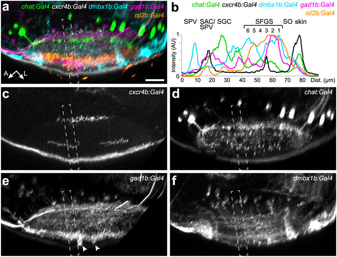
New transgenic lines label distinct sublaminae in the tectal neuropil. Lateral view of the tectal neuropil shows registered expression pattern of chat:Gal4, cxcr4b:Gal4, dmbx1b:Gal4, gad1b:Gal4, and isl2b:Gal4, as merged (a) or single channels (c?f). (b) Fluorescence intensity plots along the boxed regions in (a?f). Intensity peaks of isl2b:Gal4 expression were used for layer determination. SIN cell bodies labeled by gad1b:Gal4 are marked by arrowheads in (e). The peak for dmbx1b:Gal4 in the SPV layer reflects labeled periventricular cell bodies.
Acknowledgments
This image is the copyrighted work of the attributed author or publisher, and
ZFIN has permission only to display this image to its users.
Additional permissions should be obtained from the applicable author or publisher of the image.
Full text @ Sci. Rep.

