
|
Fig. 4
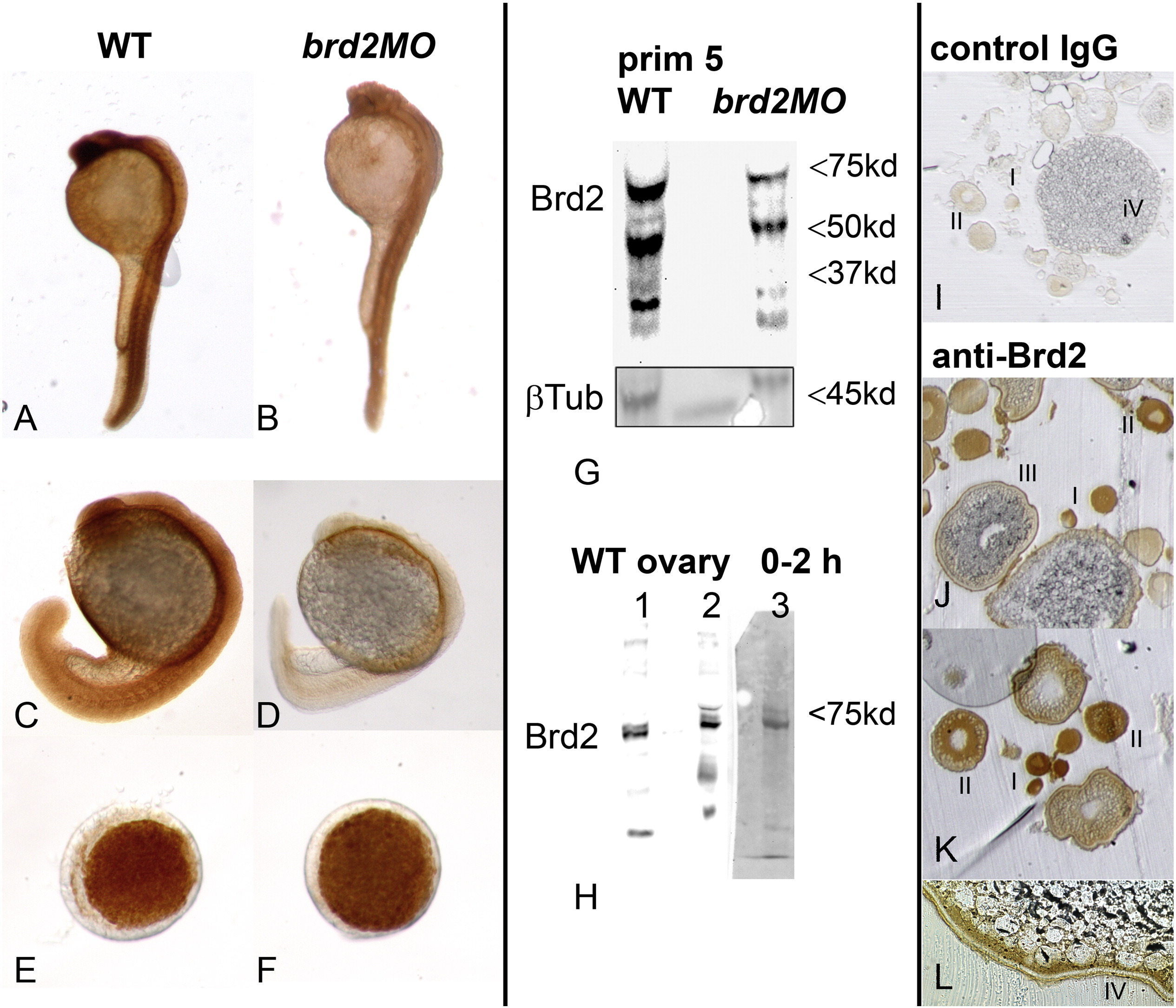
Brd2a protein is reduced in segmentation stage morphants, while maternal protein may persist in early embryos. Levels of Brd2a protein were assessed in uninjected (WT) and 8 ng brd2aMO1-injected (brd2aMO) embryos by immunohistochemistry (A?F wholemounts; I?L sections) and western blot (G, H) using the peptide antibody raised against zebrafish Brd2a. Morphant embryos at prim 5 (A,B) and 18 hpf (B,D) show reduced levels of Brd2a compared to wildtype uninjected embryos (A,C), while early embryos at 4 hpf show equivalent levels (E, F; animal pole view). Western blot shows a major protein band near 75 kD for Brd2a in prim 5 embryos (G), ovaries (H; lane 1, whole ovary; lane 2, stage I-II oocytes), and 0?2 hpf embryos (H; lane 3). Minor bands > 80 kD are often present in ovaries and early embryos, and sometimes prim 5. Putative Brd2a degradation products (or isoforms) near 55 and 37 kD are present in most preparations of prim 5 embryos (G). Reduced levels of Brd2a in brd2aMO1 prim 5 embryos are seen compared to wildtype uninjected controls (G: WT vs. brd2aMO), consistent with immunohistochemical data. Blot was reprobed for ?-tubulin as loading control. Brd2a protein is detected in both unfertilized oocytes and 0?2 hpf stage embryos (H; sectioned tissue in J?L), and becomes localized to the cortical periphery of stage III-IV oocytes, tracking with its cognate mRNA (J?L). Negative control IgG in place of anti-Brd2a shows only background staining in tissue sections (I).
Reprinted from Mechanisms of Development, 146, Murphy, T., Melville, H., Fradkin, E., Bistany, G., Branigan, G., Olsen, K., Comstock, C.R., Hanby, H., Garbade, E., DiBenedetto, A.J., Knockdown of epigenetic transcriptional co-regulator Brd2a disrupts apoptosis and proper formation of hindbrain and midbrain-hindbrain boundary (MHB) region in zebrafish, 10-30, Copyright (2017) with permission from Elsevier. Full text @ Mech. Dev.