
|
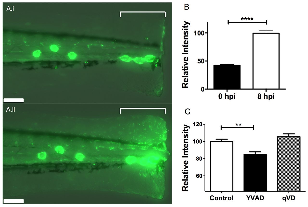
Fig. 3
Caspase-1 inhibitor YVAD downregulates NF-κB activation in response to injury. (A) Fluorescence photomicrograph of pNF-κB:EGFP embryos following tailfin transection at 1 hpi (i) and 8 hpi (ii) indicating the region quantified (square bracket). Scale bars: 100 μm. (B) There is a 2.4-fold increase in EGFP fluorescence in response to injury, quantifiable as average fluorescent intensity across the transection site (indicated in A). ****Pd0.0001 by t-test. (C) Embryos treated with 50 μM YVAD show a reduction in EGFP fluorescence at 8 hpi compared with DMSO-treated controls and embryos treated with qVD. **Pd0.01 by one-way ANOVA with Dunnett?s post-test. n=30 performed as three independent experiments.