
|
Fig. 1
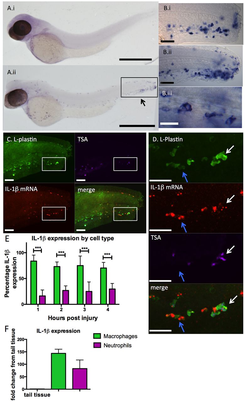
IL-1β expression is induced in leukocytes throughout the embryo in response to injury. Expression analysis of IL-1β by in situ hybridisation. (Ai) Embryos fixed at 48 hours post-fertilisation (hpf) show no IL-1β expression before injury, but IL-1β expression can be detected in cells throughout the embryo 2 hours post injury (hpi) by tailfin transection (Aii). Arrow indicates area represented in Bi,ii. Scale bars: 500 μm. (Bi?ii) IL-1β expression at the site of injury appears localised to cells with typical leukocyte morphology: close up views of region represented in Aii by a box in (Bi) 24 hpf embryos at 2 hpi and (Bii) 48 hpf embryos 2 hpi. Scale bars: 50 μm. (Biii) Magnified image of tail region: IL-1β-positive cells have large nuclei and leukocytic morphology. Scale bar: 20 μm. (C) 48-hpf embryos were fixed and at 2 hpi were probed with anti-L-plastin (green; labelling leukocytes), stained for endogenous neutrophil peroxidase activity with Cy5 TSA (purple) and FISH performed to detect IL-1β mRNA (red) to determine the localisation of IL-1β in response to injury. Scale bars: 40 μm. (D) Close-up of boxed area shown in C. Scale bars: 40 μm. IL-1β was detected both in neutrophils (TSA+;L-plastin+, white arrows) and macrophages (TSA;L-plastin+, blue arrows). (E) Quantification of IL-1β-expressing cells revealed expression predominantly in macrophages at all time points assayed (***Pd0.001 by multiple t-test with Bonferroni correction, n=8 performed as two independent experiments). (F) IL-1β was detected at very high levels by qRT-PCR in FACS-isolated zebrafish leukocytes when normalised to uninjured tail tissue.