
|
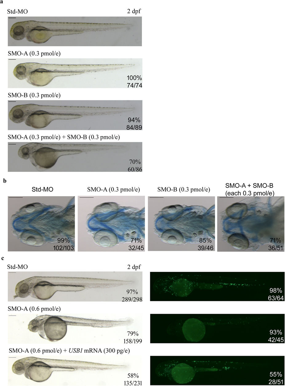
Fig. 6
Phenotypes of morphants co-injected with SMO-A and SMO-B at subphenotypic dosage and rescue of morpholino-induced phenotypes with human USB1 RNA.
(a) Representative pictures of zebrafish embryos injected with Std-MO (0.6?pmol/embryo), SMO-A (0.3?pmol/embryo), SMO-B (0.3 pmol/embryo) and co-injected with SMO-A and SMO-B (each at 0.3?pmol/embryo). (b) Alcian blue staining at 5?dpf highlights the regular morphologic architecture of the pharyngeal arch cartilages in embryos injected with Std-MO, SMO-A and SMO-B at sub-phenotypic dosages and the aberrant cartilaginous structures in embryos co-injected with SMO-A and SMO-B (each at 0.3?pmol/embryo). (c) In the left panels, lateral views of 2?dpf embryos injected with Std-MO, SMO-A (0.6?pmol/embryo) and SMO-A and human USB1 RNA (300?pg/embryo). In the right panels, fluorescent images of tg(mpx:GFP;lyzC:dsRED) embryos at 2?dpf. The green signals, representing mpx-expressing cells, are reduced in embryos injected with SMO-A (0.6?pmol/embryo) as compared to the controls, but are enhanced in embryos co-injected with human USB1 RNA (300?pg/embryo). The fractions of embryos exhibiting the investigated phenotypes out of the total number of those examined are indicated. All images are at the same magnification. Scale bars: 200??m.