
|
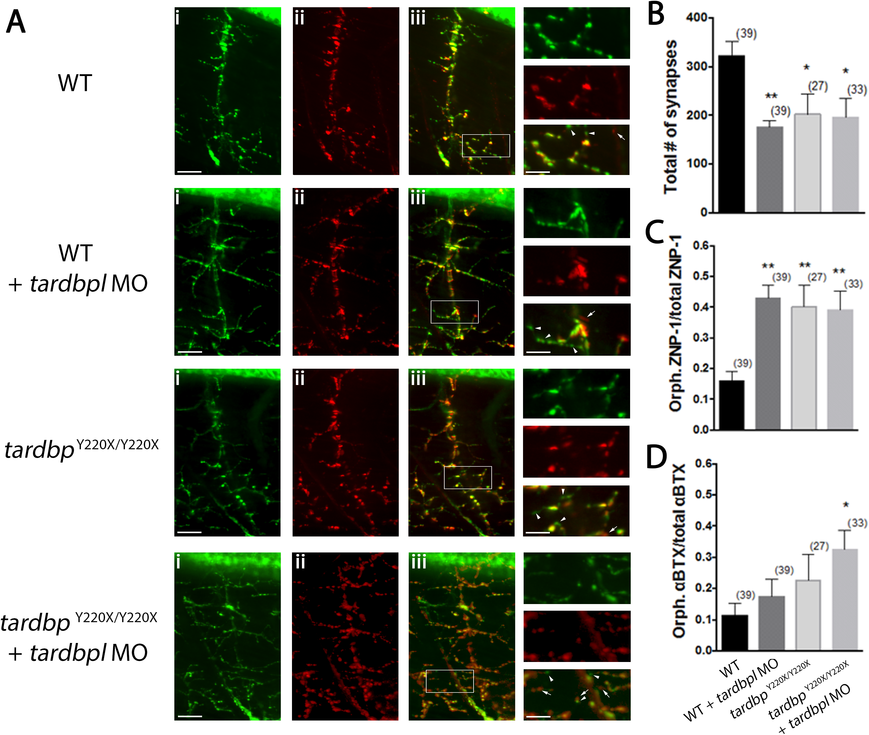
Fig. 5
Motor neuron projections in tardbpY220X/Y220X + MO larvae show increased number of orphaned presynaptic and postsynaptic puncta.
A, Representative images of single ventral root projection double-labelled for ZNP-1 (presynaptic marker, i) and sulforhodamine-conjugated ?BTX (postsynaptic marker, ii). WT larvae display extensive co-localization of both ZNP-1 and ?BTX (merged image, iii). Scale bar in (iii) and insets represent 25 ?m and 10 ?m respectively, arrowheads indicate orphaned ZNP-1 puncta and arrows indicate orphaned ?BTX labelling. B, Quantification of the number synapses formed, quantified as the number of colocalized ZNP-1 and ?BTX puncta. WT + MO (p < 0.05), tardbpY220X/Y220X (p < 0.01) and tardbpY220X/Y220X + MO larvae (p < 0.01) all had a significantly reduced number of synapses at the NMJ compared to WT larvae. C, Quantification of orphaned presynaptic ZNP-1 puncta over total number of ZNP-1 puncta. WT + MO, tardbpY220X/Y220X and tardbpY220X/Y220X + MO larvae displayed significantly higher proportion of orphaned ZNP-1 puncta compared to WT animals (p < 0.01). D, Quantification of orphaned postsynaptic ?BTX staining over total number of ?BTX puncta. tardbpY220X/Y220X + MO fish displayed significantly higher proportion of orphaned ?BTX puncta compared to WT larvae (p < 0.05). Numbers in parentheses represent the number of somites analyzed for each treatment group. Data expressed as mean � SEM: *p < 0.05; **p < 0.01.