
|
Fig. 3
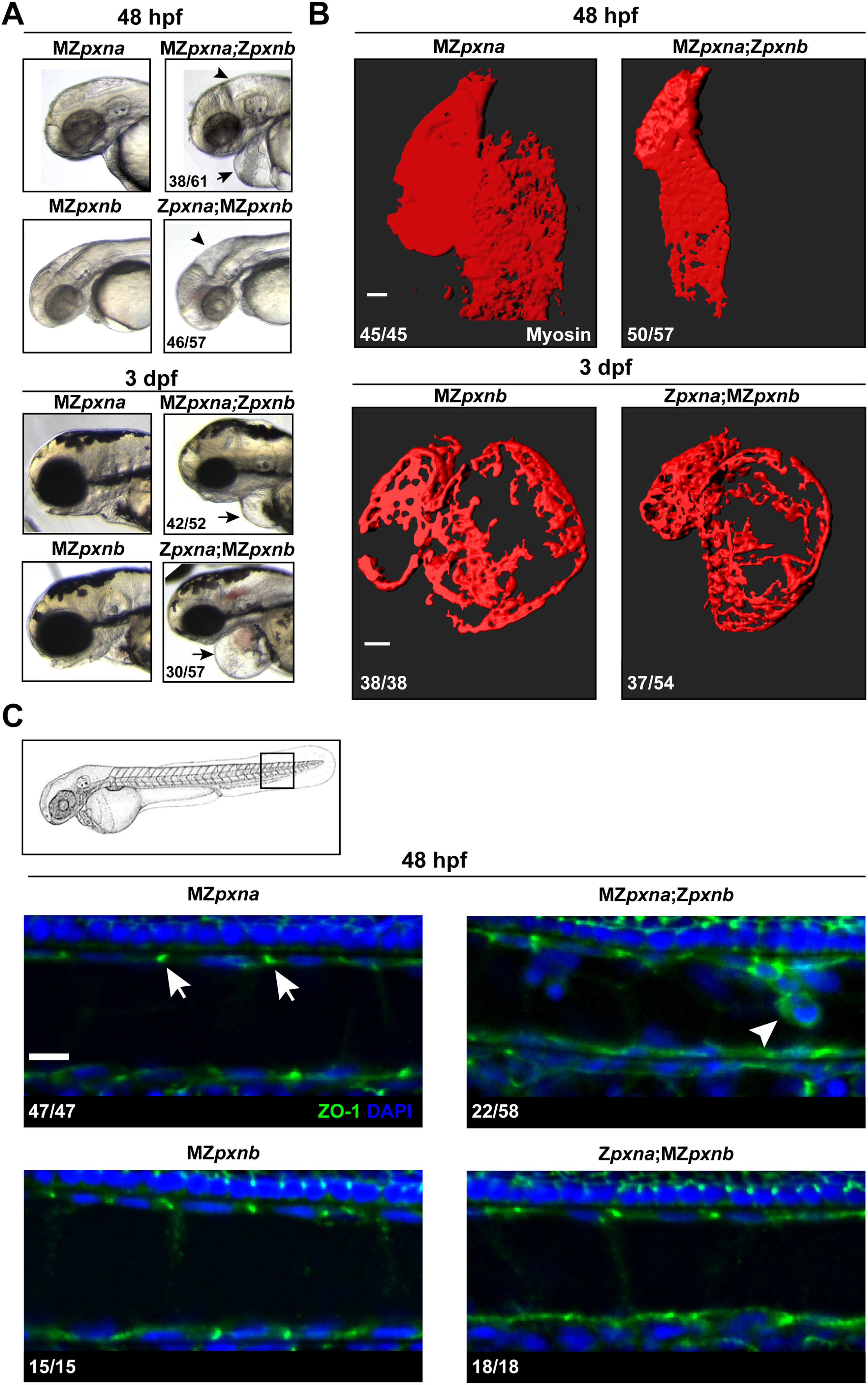
Maternal Paxillin expression differentially regulates cardiovascular and notochord development. (A) Gross morphological examination of the head and pericardial regions revealed that pxn double mutant embryos lacking maternal pxna expression (MZpxna;Zpxnb) exhibited swelling near the brain (arrowhead) and heart (arrow) at an earlier time point than those lacking maternal pxnb (Zpxna;MZpxnb). The number of MZ double mutants exhibiting the depicted phenotype over the total number of double mutants analyzed for each stage is shown. (B) 3D surface projections of Myosin immunostaining of the developing heart at 48 hpf or 3 dpf in MZpxna;Zpxnb and Zpxna;MZpxnb embryos. Disrupted cardiac morphology was observed in both pxn MZ double mutant genotypes. The number of hearts observed with the depicted phenotypes over the total number of embryos analyzed for each genotype is shown. Scale bars=50 µm. (C) At 48 hpf, outer notochord sheath cells in the tail of the embryo (boxed region of embryo diagram) normally exhibit punctate ZO-1 distribution at cell-cell junctions in sagittal optical sections (arrows) as observed in MZpxna, MZpxnb and Zpxna:MZpxnb embryos. A portion of MZpxna;Zpxnb embryos exhibited sheath cell rounding and extrusion from the monolayer with disrupted ZO-1 distributions (arrowhead). The number of notochords observed with the depicted phenotypes over the total number of embryos analyzed for each genotype is shown. Scale bar=10 µm.
Reprinted from Developmental Biology, 425(1), Jacob, A.E., Amack, J.D., Turner, C.E., Paxillin Genes and Actomyosin Contractility Regulate Myotome Morphogenesis in Zebrafish, 70-84, Copyright (2017) with permission from Elsevier. Full text @ Dev. Biol.