
|
Fig. 7
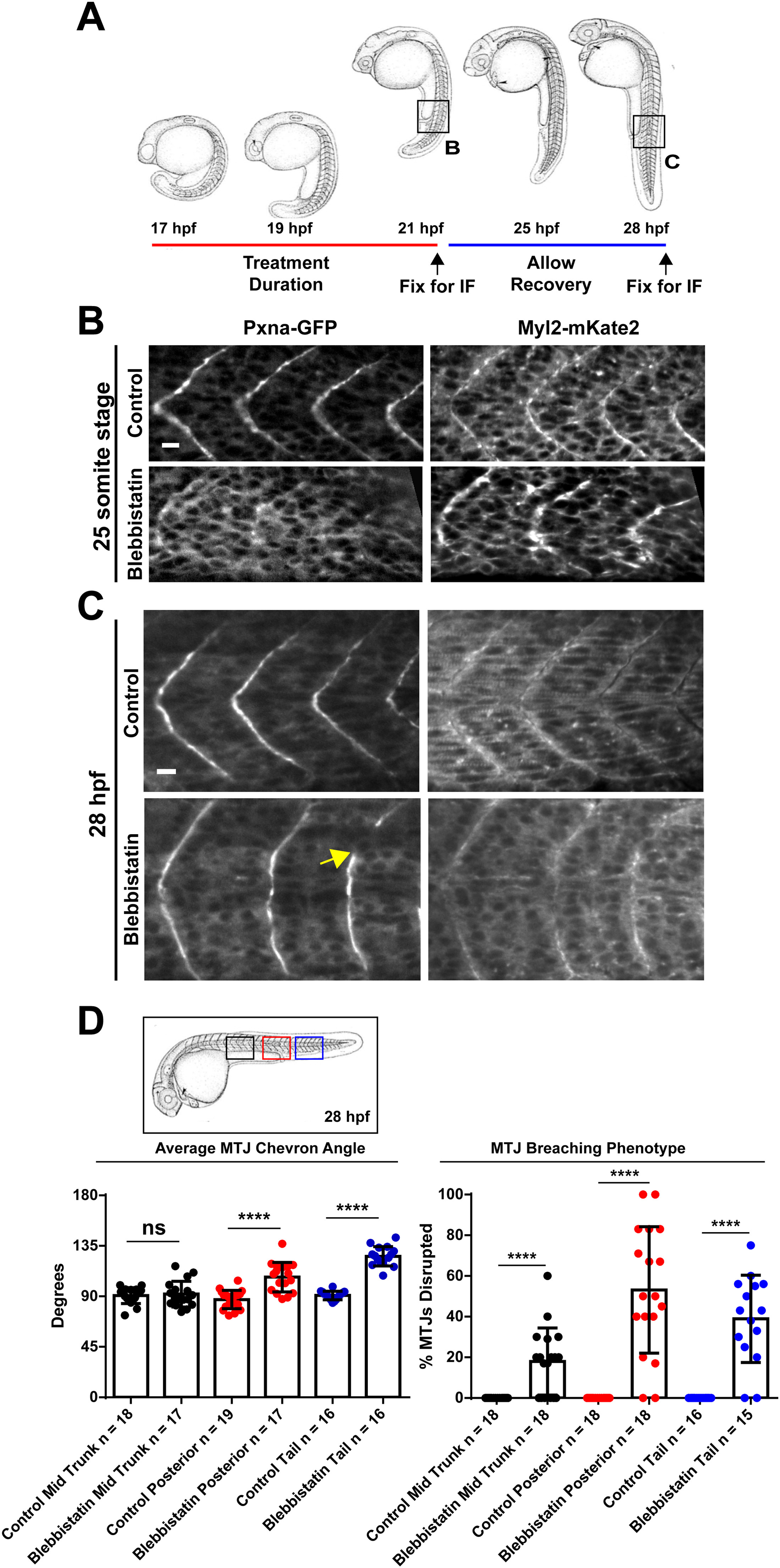
NMM-II Activity Shapes the Developing Myotome. (A) Diagram depicting the timing of pharmacological treatments with blebbistatin to inhibit NMM-II and the immunofluorescence (IF) staining of MTJ markers. Regions visualized by IF are boxed. (B) Embryos treated with blebbistatin formed MTJs marked by Myl12.1-mKate2, but Pxna-GFP MTJ localization was reduced as compared to control embryos at the 25 somite stage (C) Embryos treated with blebbistatin that were washed and allowed to develop without the drug recovered Pxna-GFP localization to MTJs by 28 hpf. However, wider chevron angle and MTJ gaps (yellow arrow) were observed. (D) Width of MTJ angle and gaps in MTJs were quantified in blebbistatin treated and control embryos at 28 hpf in the anterior region of the trunk, the posterior region of the trunk and the tail (see boxed region of embryo diagram). Data points represent individual embryo means and horizontal bars show means for each complete dataset from three independent experiments. **** p<0.001, ns = not significant. Scale bars =25 �m.
Reprinted from Developmental Biology, 425(1), Jacob, A.E., Amack, J.D., Turner, C.E., Paxillin Genes and Actomyosin Contractility Regulate Myotome Morphogenesis in Zebrafish, 70-84, Copyright (2017) with permission from Elsevier. Full text @ Dev. Biol.