
Image
|
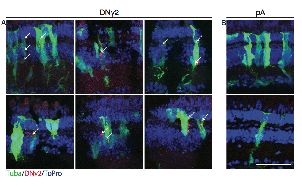
Figure Caption
Fig. S7
Expression of DN?2 in proliferating 54 cells, related to Figure 5.
Tg(tuba1a:GFP) fish were electroporated with a construct containing either a GFAP:mCh-DN?2 or GFAP:mCh-pA and allowed to recover for 96 hours, after which retinas were stained for GFP and mCherry to measure co-localization. Representative images of DN?2 (A) and pA (B) are small portions of entire retinas. Arrows indicate co-localization of mCh and GFP. Scale bar is 100?m.
Acknowledgments
This image is the copyrighted work of the attributed author or publisher, and
ZFIN has permission only to display this image to its users.
Additional permissions should be obtained from the applicable author or publisher of the image.
Full text @ Stem Cell Reports