Fig. 7
Yap1 Is Required for Vessel Maintenance
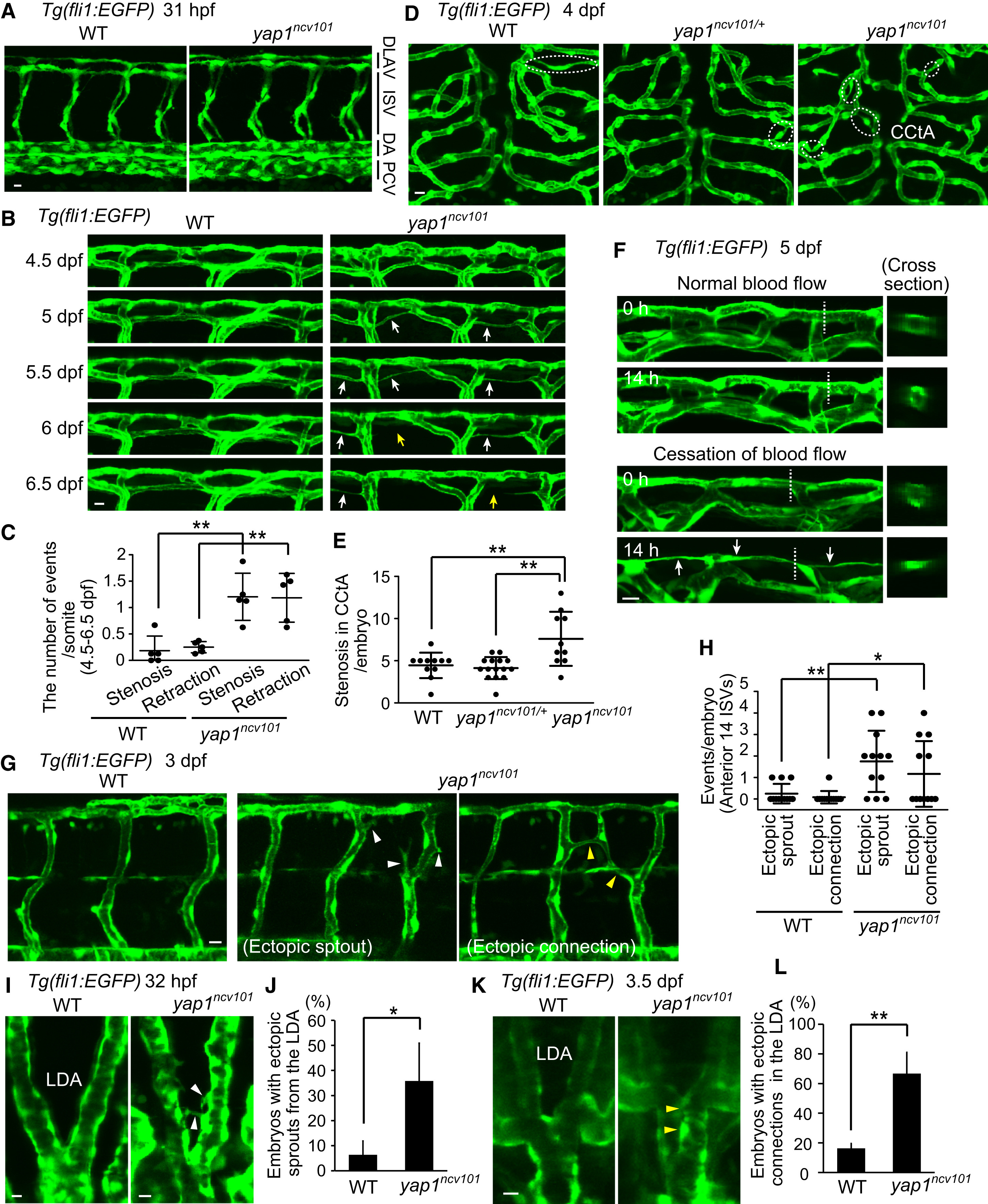
(A) Projection view of confocal stack fluorescence images of Tg(fli1:EGFP) WT (left) embryos with WT allele (left) and yap1ncv101 mutant allele (right) fixed at 31 hpf. Note the overall normal vascular formation in homozygous mutants.
(B) Projection view of confocal images of the DLAVs in Tg(fli1:EGFP) larvae with WT (left) and yap1ncv101 allele (right) taken every 12 hr from 4.5 dpf to 6.5 dpf. White and yellow arrows indicate blood vessels exhibiting lumen stenosis and EC retraction, respectively.
(C) Quantitative analyses of incidence of the DLAVs showing lumen stenosis and EC retraction at a single somite level from 4.5 to 6.5 dpf as in (B). Each dot represents the value for an embryo. For each embryo, the number of the events at six to eight somite levels were counted and then divided by the total number of somite levels observed. Horizontal lines represent mean ± SD.
(D) Projection view of confocal images of the hindbrain region in Tg(fli1:EGFP) larvae with WT allele (left), heterozygous yap1ncv101/+ allele (middle), and yap1ncv101 allele (right) at 4 dpf. Dorsal view. Dashed circles indicate lumen stenosis in the CCtA.
(E) Quantification of the number of blood vessels showing lumen stenosis in the CCtA of WT, yap1ncv101/+, or yap1ncv101 larvae at 4 dpf as observed in (D). Each dot represents the number of stenoses found in an embryo. Horizontal lines represent mean ± SD (n ? 10 independent experiments).
(F) Projection view of confocal stack fluorescence images of the DLAVs in Tg(fli1:EGFP) larvae (5 dpf) at the indicated time after cessation of blood flow by BDM treatment or without treatment. Transverse sections at the dashed lines are shown to the right. Arrows indicate blood vessels showing lumen stenosis.
(G) Projection view of confocal images of the anterior trunk ISVs in Tg(fli1:EGFP) larvae with WT allele (left) and yap1ncv101 allele (right) at 3 dpf. White and yellow arrowheads indicate ISVs exhibiting ectopic sprouts and ectopic connections, respectively.
(H) Quantification of the number of ectopic sprouts from the ISVs and ectopic connections in the ISVs within 14 ISVs of anterior trunk for each embryo at 3 dpf as in (G). Each dot represents the value for an embryo. Horizontal lines represent mean ± SD (n = 12 independent experiments).
(I) Projection view of confocal images of the LDA in Tg(fli1:EGFP) embryos with WT allele (left) and yap1ncv101 allele (right) at 32 hpf. Dorsal view. White arrowheads indicate ectopic sprouts from the LDA.
(J) Graph shows the percentage of the embryos with ectopic sprouts from the LDA at 31?32 hpf as in (I). Data are mean ± SD (n = 3 independent experiments, in each of which ?9 embryos were measured).
(K) Projection view of confocal images of the LDA in Tg(fli1:EGFP) larvae with WT allele (left) and yap1ncv101 allele (right) at 3.5 dpf. Dorsal view. Yellow arrowheads indicate ectopic connections in the LDA.
(L) Graph shows the percentage of the embryos with ectopic connections in the LDA at 3.5 dpf as in (K). Data are mean ± SD (n = 3 independent experiments, in each of which >8 embryos were measured).
?p < 0.05, ??p < 0.01. Scale bars, 10 ?m. See also Figure S7.
Reprinted from Developmental Cell, 40, Nakajima, H., Yamamoto, K., Agarwala, S., Terai, K., Fukui, H., Fukuhara, S., Ando, K., Miyazaki, T., Yokota, Y., Schmelzer, E., Belting, H.G., Affolter, M., Lecaudey, V., Mochizuki, N., Flow-Dependent Endothelial YAP Regulation Contributes to Vessel Maintenance, 523-536.e6, Copyright (2017) with permission from Elsevier. Full text @ Dev. Cell

