
|
Fig. S6
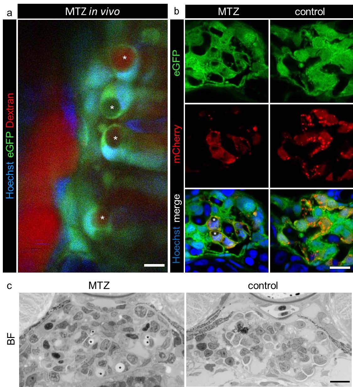
Picture a shows a 2-PM of a Chet larva which was pre-treated with MTZ for 3 hours. A few pseudocysts (asterisks) can be distinguished with different dextran fluorescence intensities (red, scale bar represents 10 ?m). Panel b shows the control confocal laser scanning micrographs to Figure 4 b. Compared to MTZ treated larvae, controls did not show pseudocysts and normal glomerular morphology (scale bar represents 5 ?m). The pictures of the methylene blue stained semithin sections in panel c confirm the appearance of these pseudocysts, as multiple encapsulated holes (asterisks) could be distinguished in MTZ treated larvae, whereas control glomeruli looked normally (scale bar represents 10 ?m).