
|
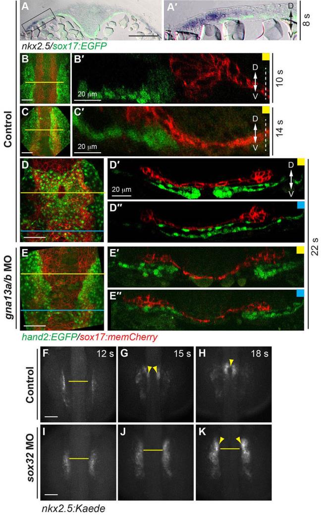
Fig. S7
The ALPM engages in subduction, with some cells migrating from the dorsal to the ventral side of the endoderm. (A) The relative positions of the ALPM and endoderm. (A-A') Overlays of GFP (endoderm) and hand 2 expression as detected by ISH in Tg(sox17:EGFP) embryos at 8s. (A'): high-magnification image of area indicated in A. (B-E'') Confocal images of control and gna13a/b MO-injected Tg(hand2:EGFP)/Tg(sox17:memCherry) embryos at the indicated stages, showing the EGFP-expressing ALPM and memCherry-expressing endoderm. (B-E) Z-projections. Horizontal lines: positions of the XZ sections. (B'-E'') XZ views of z-stacks of embryos at positions indicated in B-E. White dashed lines: midline. D: dorsal; V: ventral. (F-K) Snapshots of myocardial precursors from epifluorescence time-lapse movies taken of control (F-H) or sox32 MO-injected (I-K) Tg(nkx2.5:Kaede) embryos at the indicated stages, using a 5x/NA 0.15 objective. Yellow arrowheads: myocardial cells; yellow lines: the distance between the two populations of myocardial cells (equivalent length). Scale bars: 100 ?m, unless stated otherwise.