
|
Fig. S5
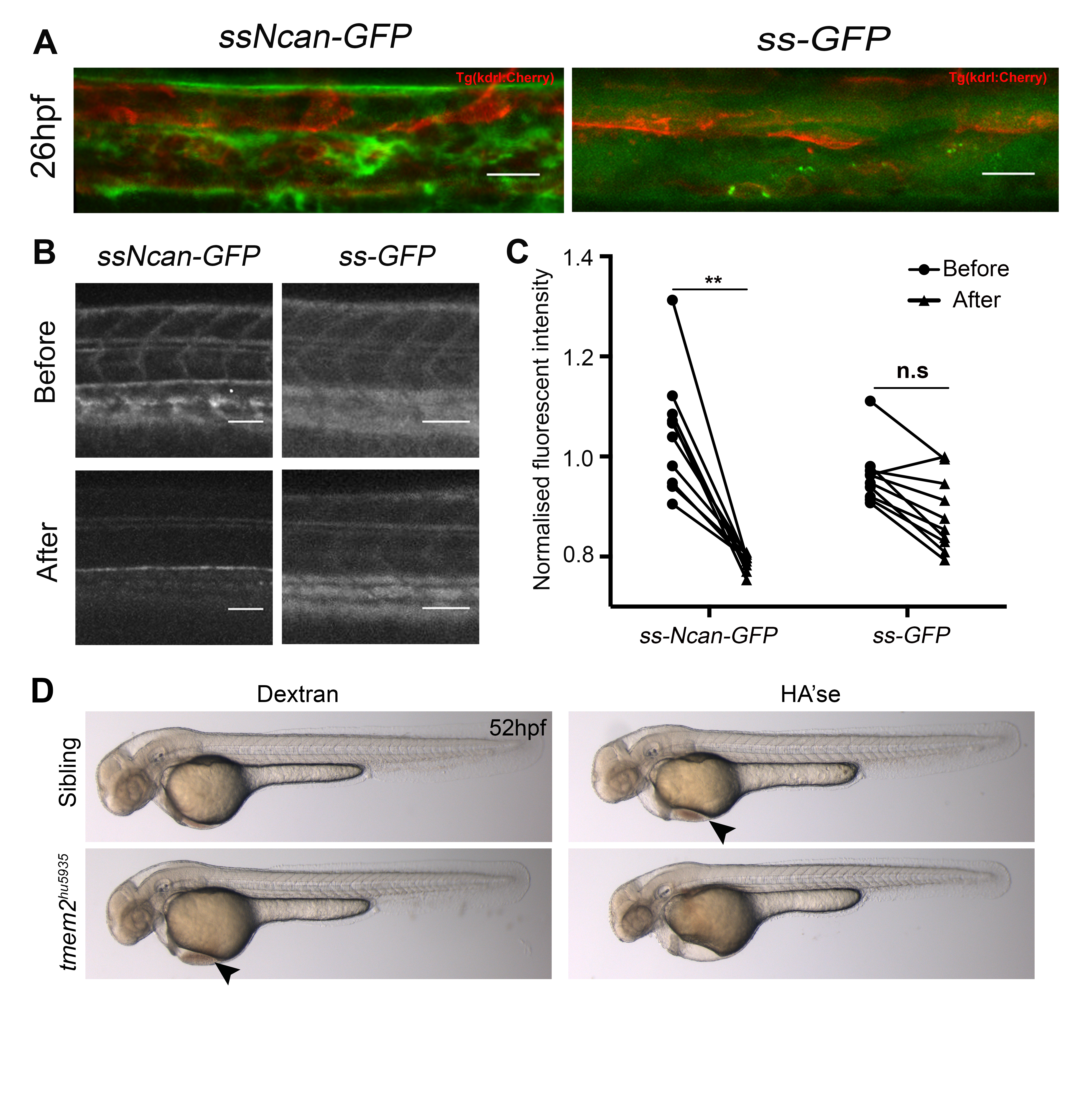
Related to Figure 3: Validation of the HA sensor and demonstration of cardiac defects following Hyaluronidase treatment
A. ssNcan-GFP localizes perivascularly whereas secreted GFP (ssGFP) disperses extracellularly and is found within the vascular lumen. Scale bar = 20 ?m. B. Degradation of the substrate, HA, by Hyaluronidase treatment. Scale bar = 50 ?m. C. A significant reduction in the fluorescence intensity of ssNcan-GFP (n=9) is observed following Hyaluronidase treatment whereas no significant difference is observed in ssGFP expressing embryos (n=10) treated with Hyaluronidase. D. Brightfield images of Dextran injected or Hyaluronidase injected sibling and mutant embryos. Hyaluronidase treatment does not rescue the cardiac defect in tmem2hu5935 mutant embryos and induces cardiac defects in sibling mutants. Mutant hearts have cardiac looping defects, resulting in pooling of blood in the pericardium at the location of the inflow tract (black arrowhead). Cardiac looping proceeds in sibling embryos injected with Hyaluronidase however blood does accumulate in the pericardium (black arrowhead; due to blood regurgitation between the chambers) and eventually results in cardiac oedema. The cardiac looping defect in tmem2 mutants injected with Hyaluronidase does not correct and cardiac oedema is more severe. ??p < 0.01.
Reprinted from Developmental Cell, 40, De Angelis, J.E., Lagendijk, A.K., Chen, H., Tromp, A., Bower, N.I., Tunny, K.A., Brooks, A.J., Bakkers, J., Francois, M., Yap, A.S., Simons, C., Wicking, C., Hogan, B.M., Smith, K.A., Tmem2 Regulates Embryonic Vegf Signaling by Controlling Hyaluronic Acid Turnover, 123-136, Copyright (2017) with permission from Elsevier. Full text @ Dev. Cell