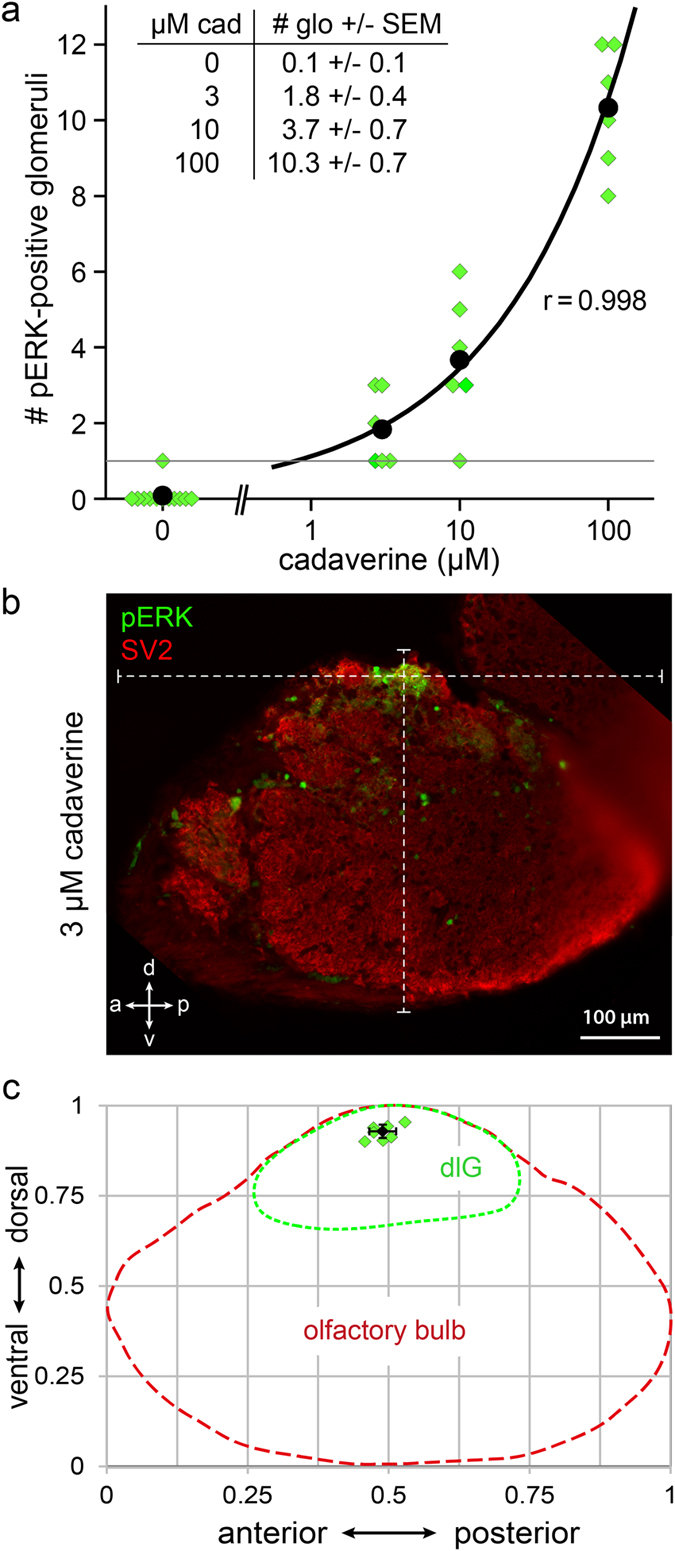
Fig. 5
A single invariant glomerulus is activated by exposure to low concentrations of cadaverine.
(a) Number of glomeruli activated in response to different doses of cadaverine was counted from sections. Water did not evoke any pERK signal in the dorsolateral cluster in all but one cases (n?=?12). At 3 and 10??M cadaverine one to few glomeruli were labelled (see table inset). At 100??M cadaverine about 10 glomeruli were labelled (n?=?6 for each cadaverine concentration used). (b) Exemplary image of a single glomerulus labelled by pERK, double labelling with SV2 to visualize all glomeruli in this section. White dashed lines intersect in the glomerulus center and show overall a?p and v?d length of the olfactory bulb section used for calculating the normalized glomerulus position. (c) Schematic representation of the dlGcad coordinates measured in the olfactory bulb. Red and green dashed lines represent olfactory bulb and dorsolateral cluster contours, respectively. Green squares represent single measurements from 7 different bulbi. Black square and error bars represent mean value?±?SD.

