
|
Fig. S1
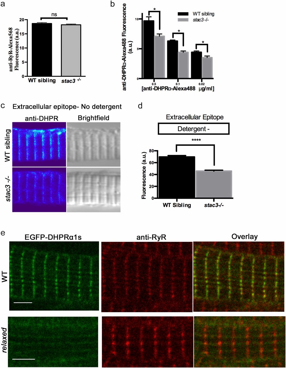
Loss of Stac3 does not prevent trafficking of DHPR? to triads. (A) Quantification of the mean of immunofluorescence labeling (�SEM) of Alexa 568 directly coupled to anti-RyR in stac3?/? at triads compared with WT siblings (n = 60, t test, P = 0.29). (B) Quantification of the mean of immunofluorescence labeling of Alexa 488 directly coupled to anti-DHPR?1S at different concentrations in stac3?/? at triads compared with WT siblings (n = 40 WT sibling, n = 40 stac3?/?, t test, *P < 0.0001). (C) Immunofluorescence (Left) and bright-field images (Right) of WT sibling and stac3?/? disassociated myotubes labeled without detergent with anti-DHPR?1S that recognizes an extracellular epitope. (D) Histogram showing that there is a decrease in T-tubule triadic DHPR?1S in stac3?/? dissociated myotubes (n = 160 WT sibling, n = 125 stac3?/?, t test, ****P < 0.0001). (E) Anti-Ryr immunolabeling of a fixed muscle fiber expressing EGFP-DHPR?1S showing that EGFP-DHPR?1S does not localize to triads in a relaxed mutant fiber, whereas it does in a WT fiber. SEMs are indicated. n.s., not significant. (Scale bars, 4 ?m.)