Fig. S12
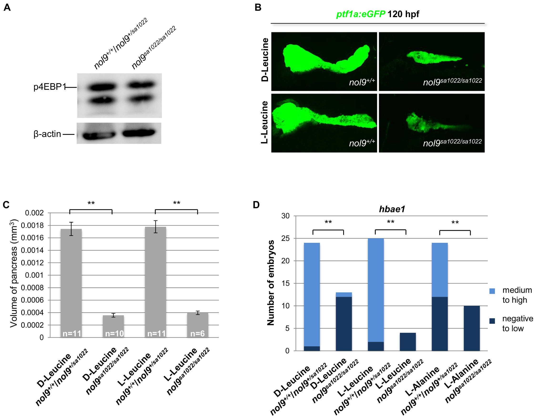
The pancreatic and hematological defects in nol9sa1022/sa1022 larvae are independent of the mTOR pathway.
(A) Western blot analysis of p4EBP1 and β-actin (loading control) in whole cell lysates of nol9sa1022/sa1022 and wt siblings at 120 hpf. (B) Representative confocal images of the pancreas of Tg(ptf1a:EGFP) nol9sa1022/sa1022 and nol9+/+ larvae at 120 hpf after treatment with L-Leucine or D-Leucine from 24 hpf. (C) The average volume of the ptf1a-positive exocrine pancreas in 120 hpf Tg(ptf1a:EGFP) larvae, depending on their genotype and treatment with either D-Leucine or L-Leucine from 24 hpf. The data are represented as the mean +/- SEM. Student’s t-test, **, p<0.01. (D) Quantification of hbae1 WISH performed on 120 hpf larvae treated with D-Leucine, L-Leucine or L-Alanine from 24 hpf. Data are represented as the number of wt (nol9+/+/nol9+/sa1022) or mutant (nol9sa1022/sa1022) larvae belonging to either phenotypic group. Fisher’s exact test, **, p<0.01.

