
Fig. 3
- ID
- ZDB-IMAGE-170208-11
- Genes
- Antibodies
- Source
- Figures for Bielczyk-Maczy?ska et al., 2015
|
Fig. 3
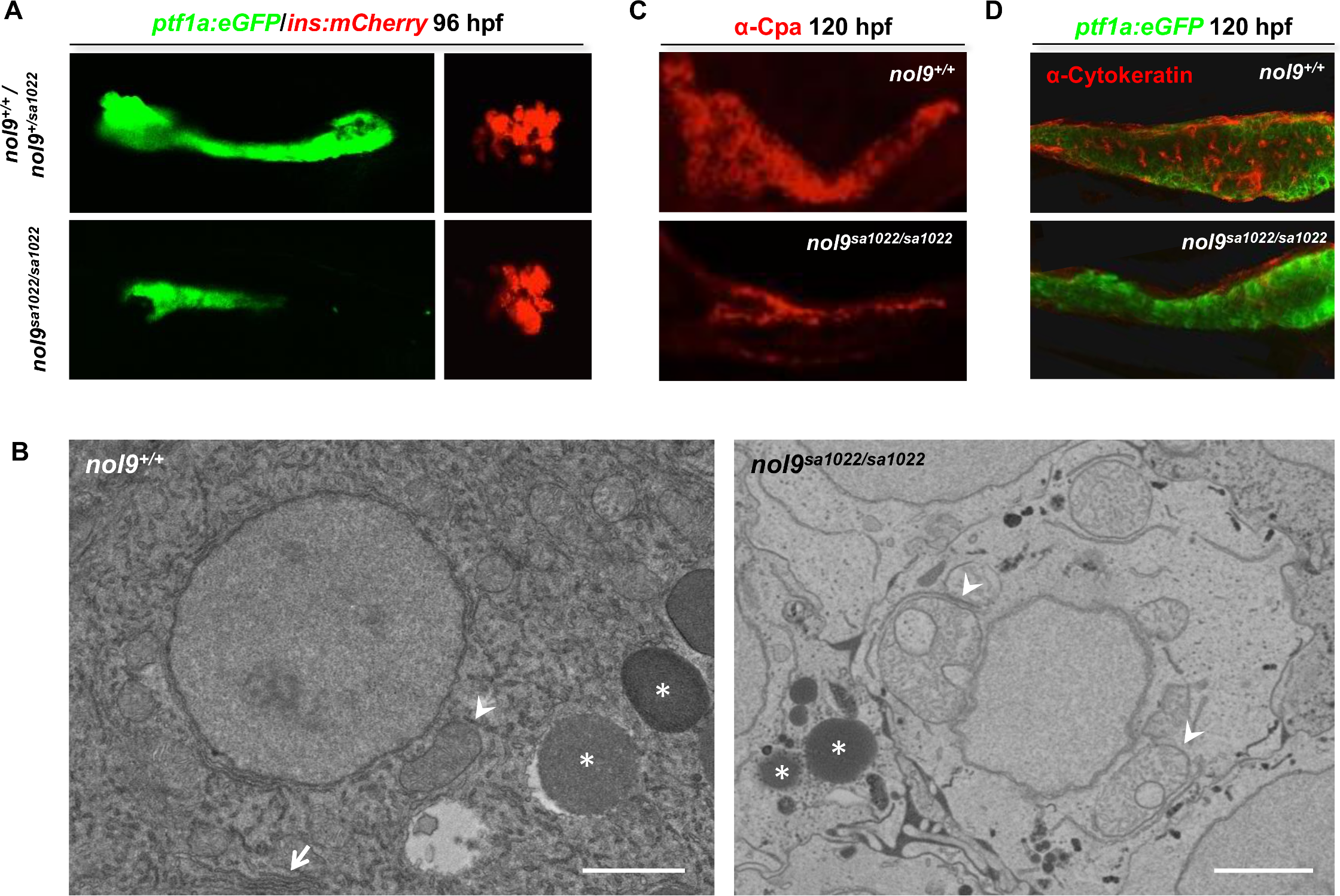
nol9 mutation affects the development of the exocrine but not the endocrine pancreas.
(A) Representative single channel confocal images of the pancreas of 96 hpf Tg(ptf1a:EGFP;ins:mCherry) larvae. nol9sa1022/sa1022 larvae were characterized by smaller ptf1a-positive area (green) and similar ins-positive area (red) compared to nol9+/+ siblings. (B) TEM pictures of the exocrine pancreas of nol9+/+ and nol9sa1022/sa1022 larvae at 120 hpf. The white arrow denotes endoplasmic reticulum in the nol9+/+ cell. Asterisks indicate zymogen granules. Arrowheads indicate mitochondria. Scale bar: 2 ?m. (C-D) Confocal images of the pancreas of Tg(ptf1a:EGFP) larvae subjected to immunohistochemistry against ?-Carboxypeptidase-a (?-Cpa) (C) and ?-Cytokeratin (D) at 120 hpf. (C) The exocrine pancreas differentiation marker ?-Cpa (red) was detected in nol9+/+ and nol9sa1022/sa1022 siblings. (D) The pancreatic ducts expressing ?-Cytokeratin (red) are not apparent in nol9sa1022/sa1022 mutants, in contrast to the ductal network clearly visible in nol9+/+ siblings.