
Image
|
Figure Caption
Fig. 2
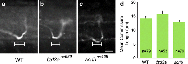
Commissure formation is not affected by loss of fzd3a or scrib. a?c Confocal micrographs showing lateral view of commissures from individual CoPA axons in WT, fzd3a rw689 , and scrib rw468 embryos. Anterior is to the left, dorsal is up, d Measurements of distance traveled by axon within the midline of the spinal cord in the anterior?posterior axis. No significant difference was found in the length of commissures in WT, fzd3a rw689 , and scrib rw468 embryos. n number of neurons counted. A two-tailed Student?s t test was used for statistical analysis. Error indicates SEM
Figure Data
Acknowledgments
This image is the copyrighted work of the attributed author or publisher, and
ZFIN has permission only to display this image to its users.
Additional permissions should be obtained from the applicable author or publisher of the image.
Full text @ BMC Neurosci.