Fig. 2
Fig. 2
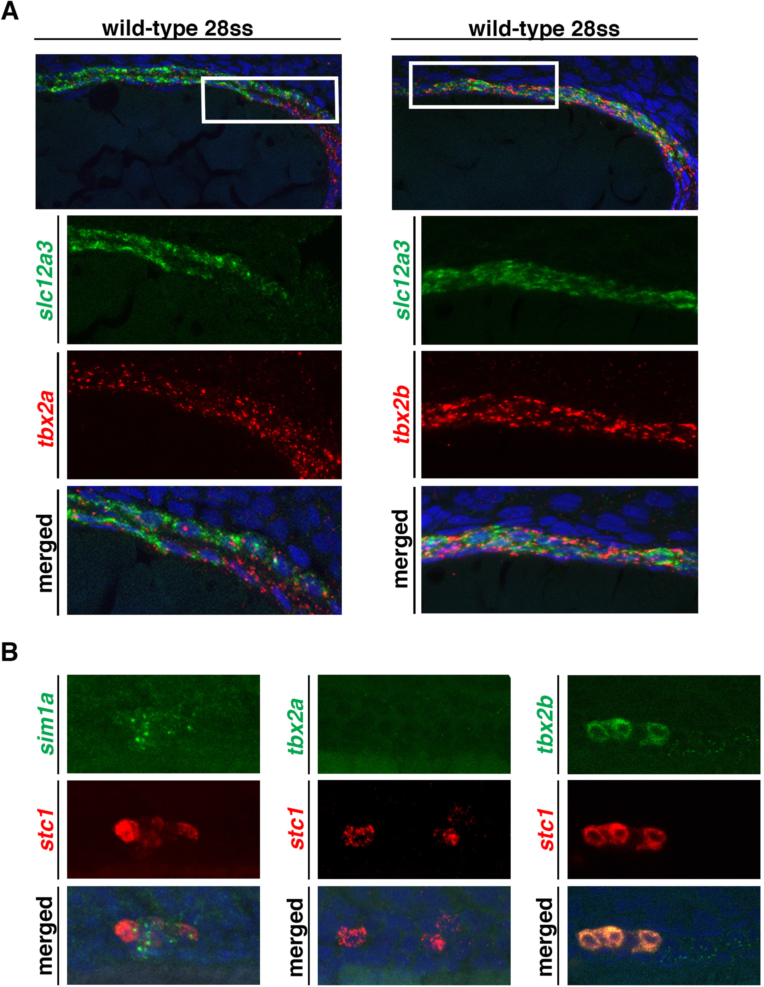
tbx2a/b exhibit co-expression with the DL and CS. (A) Fluorescent in situ hybridization was used to examine the distal region of the pronephros of wild-type 28 ss embryos, with DAPI (blue) used to label nuclei. tbx2a expression again appears strongest in the PD segment and fades as it progresses anteriorly. The region in the white box represents the moderate co-expression seen between tbx2a and slc12a3 near the boundary of the slc12a3 domain. In contrast, tbx2b expression appears strongly co-localized through most of the length of the DL, as exemplified by the region in the white box. In addition, tbx2b continues to be expressed past the DL in the PD segment. (B) The transcription factor sim1a, which has been demonstrated to be a necessary requirement for CS formation, shows specific co-localization with CS marker stc1 in wild-type, 28 ss embryos. This indicates that sim1a can be used as a secondary indicator for changes in the CS. Additionally, when examining the CS in wild-type embryos at 28 ss it was noted that tbx2a is not expressed in or near the CS while tbx2b does co-localize with stc1 in the CS at this stage.
Reprinted from Developmental Biology, 421(1), Drummond, B.E., Li, Y., Marra, A.N., Cheng, C.N., Wingert, R.A., The tbx2a/b transcription factors direct pronephros segmentation and corpuscle of Stannius formation in zebrafish, 52-66, Copyright (2017) with permission from Elsevier. Full text @ Dev. Biol.

