
|
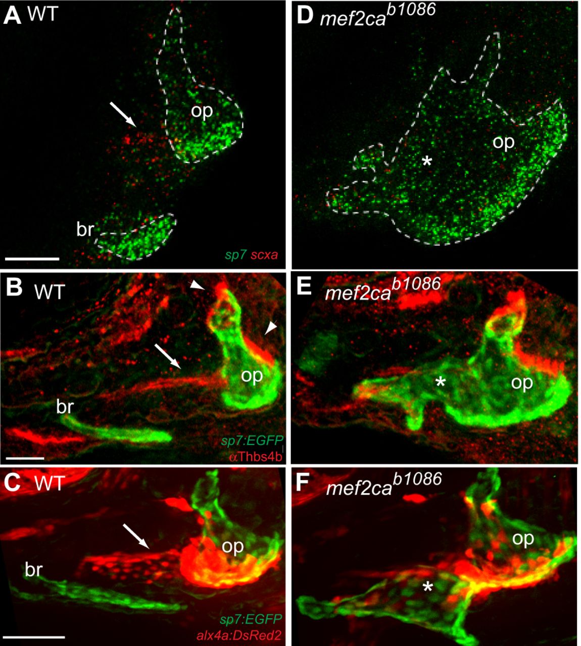
Fig. 2
The orthogonal tissue track has ligament identity, which is lost when ectopic bone develops in mef2cab1086. (A,D) Confocal images of sp7 (green) and scxa (red) transcripts detected by in situ hybridization in 5?dpf zebrafish. (B,E) Confocal images of 5?dpf sp7:EGFP (green) transgenic larvae immunostained with anti-Thbs4b antibody (red). (C,F) Confocal images of live 6?dpf sp7:EGFP (green) and alx4a:DsRed2 (red) transgenic larvae. Wild-type (WT) and mutant (mef2cab1086) images were uniformly adjusted for brightness and contrast. The opercle (op) and branchiostegal ray (br) are indicated. Arrows mark putative ligaments, arrowheads denote tendons, asterisks mark ectopic bone where ligament identity is lost. All images are projections of several confocal sections. Lateral views, anterior is towards the left and dorsal is up. Scale bars: 50?�m.