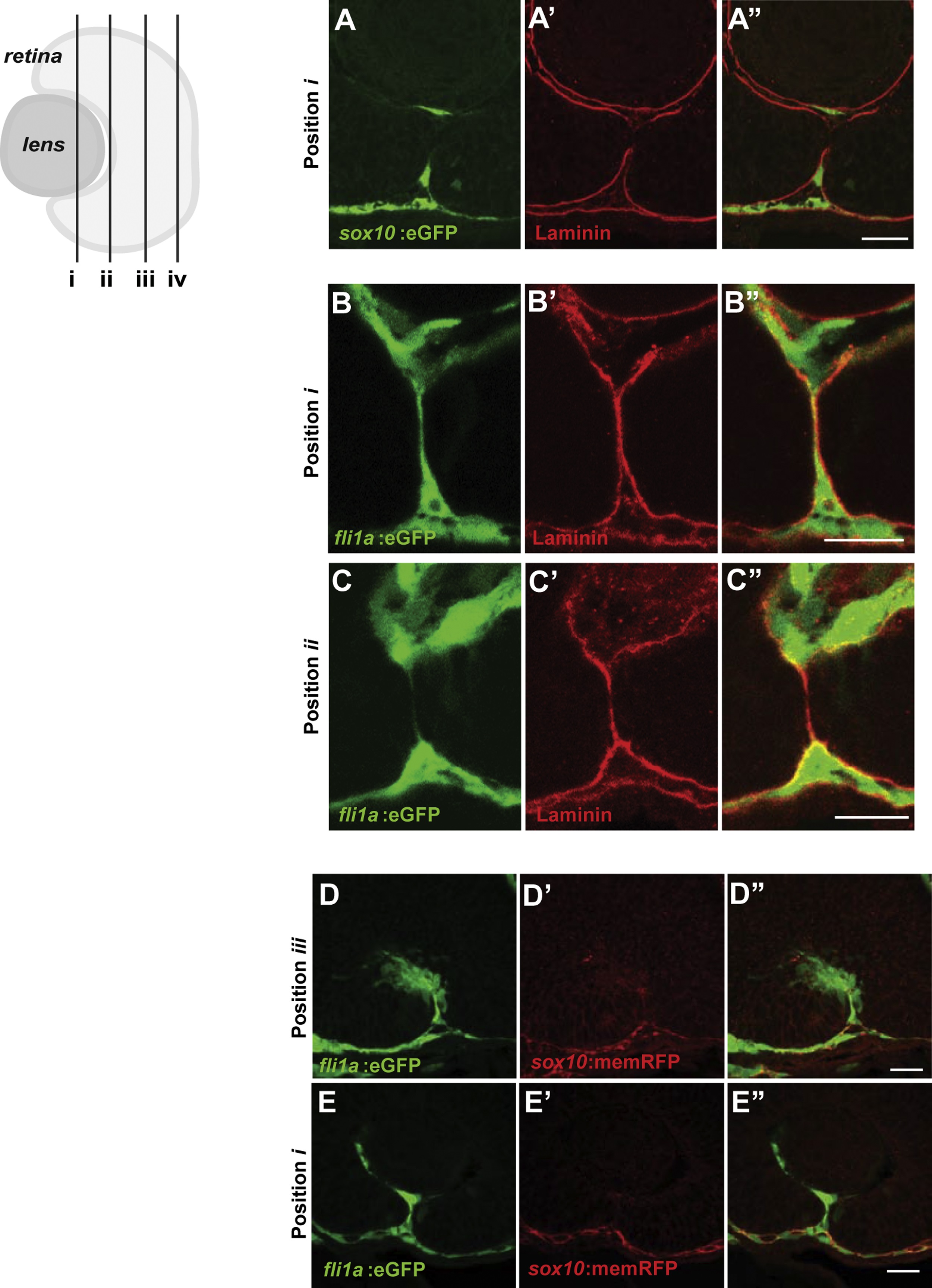
Fig. S1
Periocular mesenchyme cells migrate through the CF during CFC. (A-C) Sagittal section views of the CF immunostained with GFP and Lam-111 antibodies to visualize POM cells (green) and BM (red). Section planes as depicted in Fig. 1A. (A) sox10:eGFP cells at 37 hpf. Few sox10:eGFP cells are detected in the CF. (B,C) fli1a:eGFP cells at 36 hpf at a central and central/proximal plane within the eye. fli1a:eGFP cells spend extended durations in the CF. (D,E) Sagittal section views at central/proximal and distal/central planes of the eye showing fli1a:eGFP cells (green) and sox10:memRFP cells (red) in the CF at 34 hpf. Scale bar =20 Ám.
Reprinted from Developmental Biology, 419(2), James, A., Lee, C., Williams, A.M., Angileri, K., Lathrop, K.L., Gross, J.M., The Hyaloid Vasculature Facilitates Basement Membrane Breakdown During Choroid Fissure Closure in the Zebrafish Eye, 262-272, Copyright (2016) with permission from Elsevier. Full text @ Dev. Biol.

