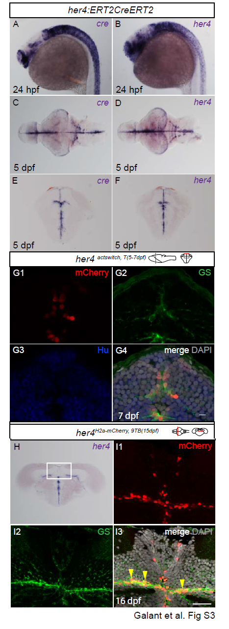
Fig. S3
Validation of the Tg(her4:ERT2CreERT2) line for faithful recombination in the post-embryonic midbrain. A-F. Compared expression of endogenous her4 and cre revealed by in situ hybridization on whole-mount embryos at 24hpf (A,B), whole-mount dissected 5dpf brains (C,D, dorsal views anterior left) and cross-sectioned 5dpf midbrains (E,F). G. Compared expression of mCherry recombined in her4actswitch,T(5-7dpf) larvae and analysed at 7dpf, with expression of the glial marker GS and the neuronal marker HuC/D. Confocal views of cross sections at the level indicated on the schematic. Note that mCherry expression is faithfully restricted to tectal RG (G4), a location identical to endogenous her4 expression (compare with F). H, I. Validation of the Tet-On approach. H. Expression of endogenous her4 revealed by in situ hybridization in a 15dpf brain. I1-3. High magnification of the area boxed in H. Compared expression of H2A-mCherry and GS in a her4H2a-mCherry,9BT(15dpf) animal analysed one day post treatment (ie at 16 dpf). Confocal views. I1-I2 are single channels (color-coded), and I3 is a merged view with DAPI counterstaining. Note the coincidence of H2A-mCherry and RG in the TeO (arrowheads in I3). Note also that induction faithfully reflects endogenous her4 expression (compare with H). Scale bars: G 5?m , I 50?m. Abbreviations: TeO: tectum opticum.
Reprinted from Developmental Biology, 420(1), Galant, S., Furlan, G., Coolen, M., Dirian, L., Foucher, I., Bally-Cuif, L., Embryonic origin and lineage hierarchies of the neural progenitor subtypes building the zebrafish adult midbrain, 120-135, Copyright (2016) with permission from Elsevier. Full text @ Dev. Biol.

